
Deposition Date
2022-07-22
Release Date
2023-01-25
Last Version Date
2024-04-03
Entry Detail
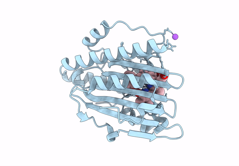
PDB ID:
7YKB
Keywords:
Title:
Neutron Structure of PcyA D105N Mutant Complexed with Biliverdin at Room Temperature
Biological Source:
Source Organism(s):
Synechocystis sp. PCC 6803 substr. Kazusa (Taxon ID: 1111708)
Expression System(s):
Method Details:
Experimental Method:
R-Value Free:
['0.17
R-Value Work:
['0.15
R-Value Observed:
['0.15
Space Group:
P 21 21 2
