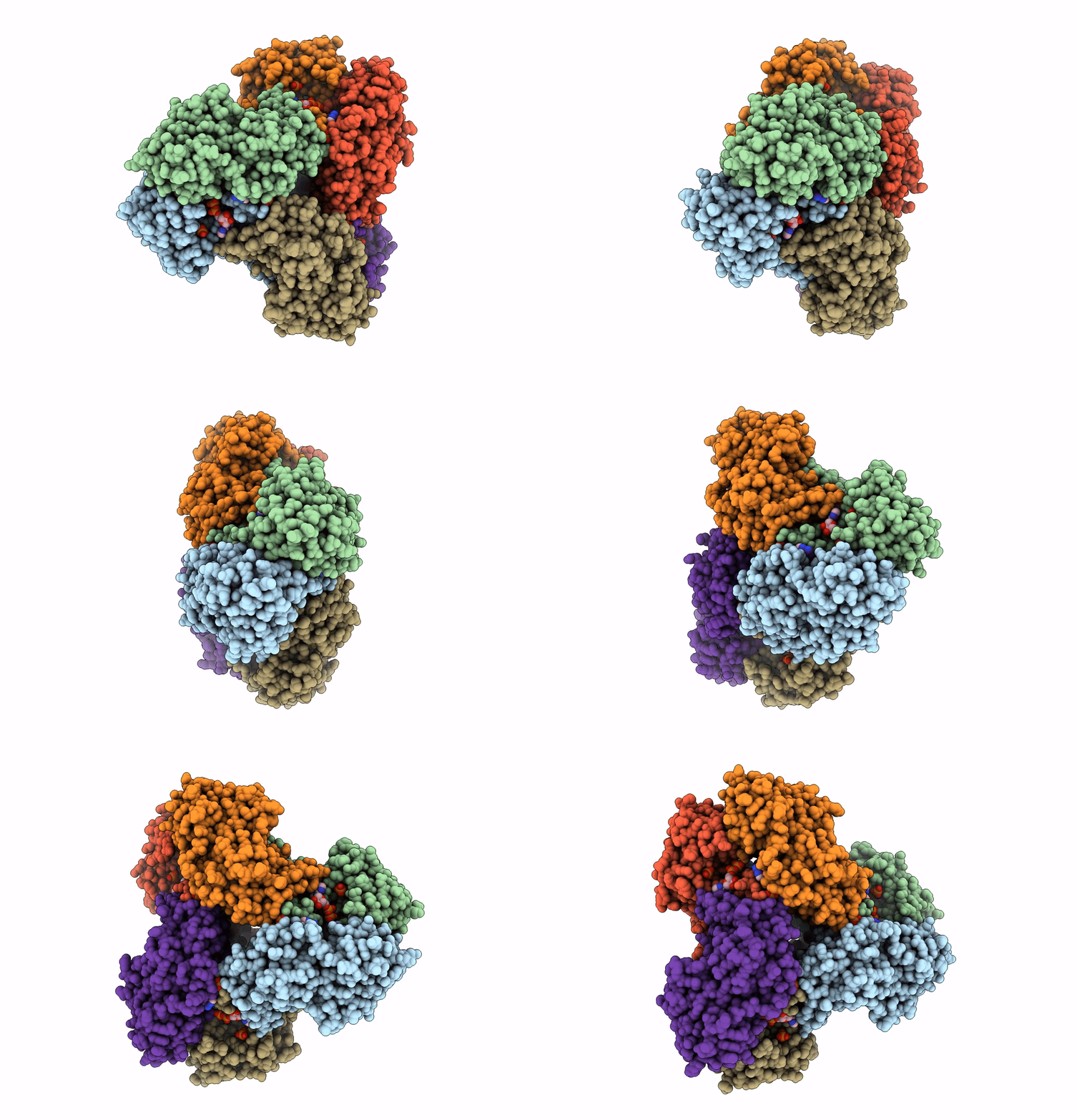
7YK1 image
Deposition Date 2022-07-21
Release Date 2023-08-02
Last Version Date 2023-09-20
Entry Detail
PDB ID:
7YK1
Keywords:
Title:
Structural basis of human PRPS2 filaments
Biological Source:
Source Organism:
Homo sapiens (Taxon ID: 9606)
Method Details:
Experimental Method:
Resolution:
3.08 Å
Aggregation State:
FILAMENT
Reconstruction Method:
SINGLE PARTICLE
Macromolecular Entities
Polymer Type:polypeptide(L)
Molecule:Ribose-phosphate pyrophosphokinase 2
Gene (Uniprot):PRPS2
Chain IDs:A, B, C, D, E, F
Chain Length:324
Number of Molecules:6
Biological Source:Homo sapiens
Primary Citation
Structural basis of human PRPS2 filaments.
Cell Biosci 13 100 100 (2023)
PMID: 37248548 DOI: 10.1186/s13578-023-01037-z

Abstact

BACKGROUND PRPP synthase (PRPS) transfers the pyrophosphate groups from ATP to ribose-5-phosphate to produce 5-phosphate ribose-1-pyrophosphate (PRPP), a key intermediate in the biosynthesis of several metabolites including nucleotides, dinucleotides and some amino acids. There are three PRPS isoforms encoded in human genome. While human PRPS1 (hPRPS1) and human PRPS2 (hPRPS2) are expressed in most tissues, human PRPS3 (hPRPS3) is exclusively expressed in testis. Although hPRPS1 and hPRPS2 share 95% sequence identity, hPRPS2 has been shown to be less sensitive to allosteric inhibition and specifically upregulated in certain cancers in the translational level. Recent studies demonstrate that PRPS can form a subcellular compartment termed the cytoophidium in multiple organisms across prokaryotes and eukaryotes. Forming filaments and cytoophidia is considered as a distinctive mechanism involving the polymerization of the protein. Previously we solved the filament structures of Escherichia coli PRPS (ecPRPS) using cryo-electron microscopy (cryo-EM) 1. RESULTS Order to investigate the function and molecular mechanism of hPRPS2 polymerization, here we solve the polymer structure of hPRPS2 at 3.08 Å resolution. hPRPS2 hexamers stack into polymers in the conditions with the allosteric/competitive inhibitor ADP. The binding modes of ADP at the canonical allosteric site and at the catalytic active site are clearly determined. A point mutation disrupting the inter-hexamer interaction prevents hPRPS2 polymerization and results in significantly reduced catalytic activity. CONCLUSION Findings suggest that the regulation of hPRPS2 polymer is distinct from ecPRPS polymer and provide new insights to the regulation of hPRPS2 with structural basis.

Legend

Protein

Chemical

Disease

Primary Citation of related structures