7YJ0 image
Deposition Date 2022-07-18
Release Date 2023-05-10
Last Version Date 2024-05-08
Entry Detail
PDB ID:
7YJ0
Keywords:
Title:
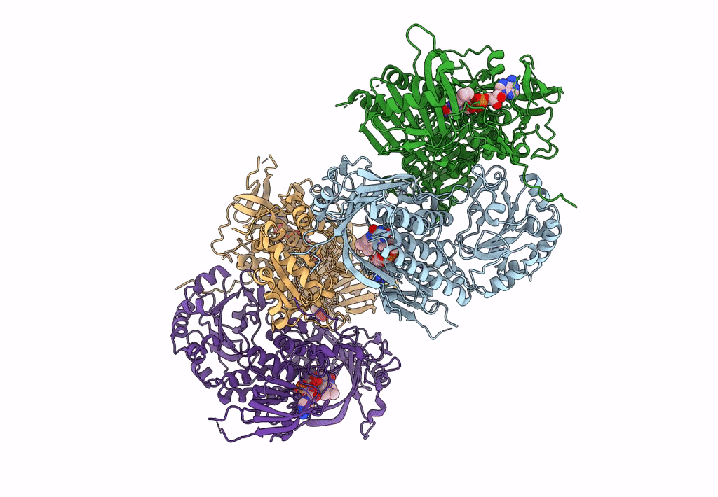
Structural basis of oxepinone formation by a flavin-monooxygenase VibO
Biological Source:
Source Organism:
Host Organism:
Method Details:
Experimental Method:
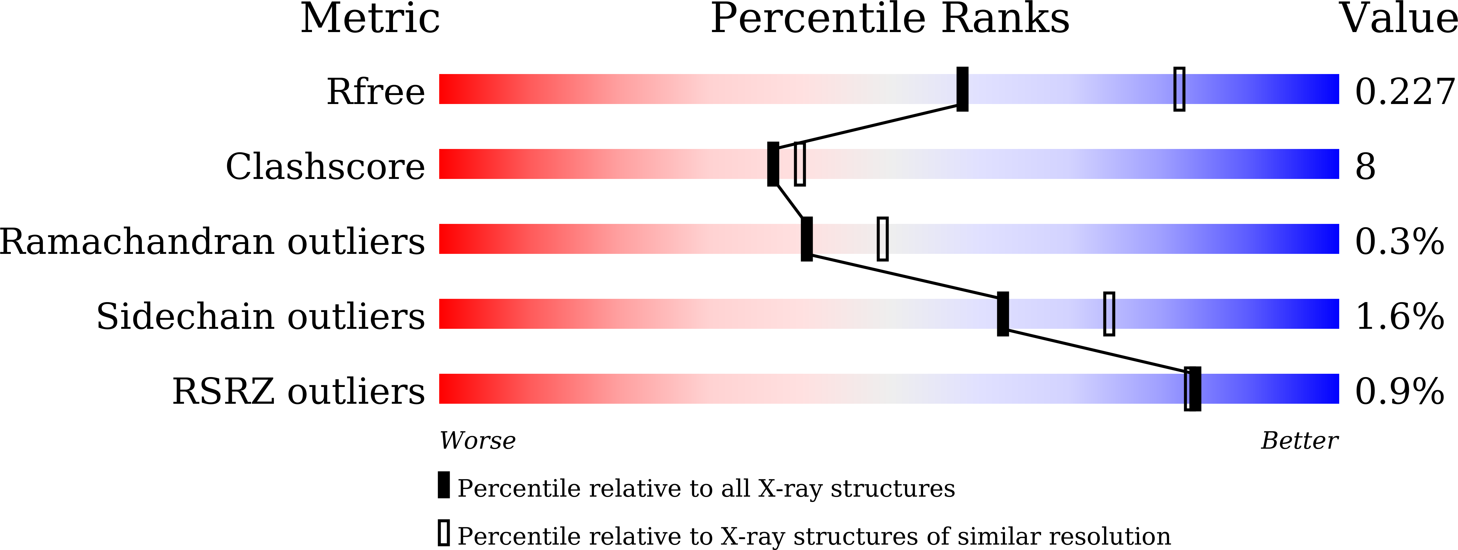
Resolution:
2.43 Å
R-Value Free:
0.22
R-Value Work:
0.17
R-Value Observed:
0.17
Space Group:
P 1
Macromolecular Entities
Structures with similar UniProt ID
Protein Blast
Polymer Type:polypeptide(L)
Molecule:VibO
Chain IDs:A, B, C, D
Chain Length:653
Number of Molecules:4
Biological Source:Boreostereum vibrans
Primary Citation
A flavin-monooxygenase catalyzing oxepinone formation and the complete biosynthesis of vibralactone.
Nat Commun 14 3436 3436 (2023)
PMID: 37301868 DOI: 10.1038/s41467-023-39108-x

Abstact

Oxepinone rings represent one of structurally unusual motifs of natural products and the biosynthesis of oxepinones is not fully understood. 1,5-Seco-vibralactone (3) features an oxepinone motif and is a stable metabolite isolated from mycelial cultures of the mushroom Boreostereum vibrans. Cyclization of 3 forms vibralactone (1) whose β-lactone-fused bicyclic core originates from 4-hydroxybenzoate, yet it remains elusive how 4-hydroxybenzoate is converted to 3 especially for the oxepinone ring construction in the biosynthesis of 1. In this work, using activity-guided fractionation together with proteomic analyses, we identify an NADPH/FAD-dependent monooxygenase VibO as the key enzyme performing a crucial ring-expansive oxygenation on the phenol ring to generate the oxepin-2-one structure of 3. The crystal structure of VibO reveals that it forms a dimeric phenol hydroxylase-like architecture featured with a unique substrate-binding pocket adjacent to the bound FAD. Computational modeling and solution studies provide insight into the likely VibO active site geometry, and suggest possible involvement of a flavin-C4a-OO(H) intermediate.

Legend

Protein

Chemical

Disease

Primary Citation of related structures