7YGG image
Deposition Date 2022-07-11
Release Date 2022-10-19
Last Version Date 2024-10-09
Entry Detail
PDB ID:
7YGG
Keywords:
Title:
Crystal structure of human CD47 in complex with engineered SIRPa.D1(N80A)
Biological Source:
Source Organism:
Homo sapiens (Taxon ID: 9606)
Host Organism:
Method Details:
Experimental Method:
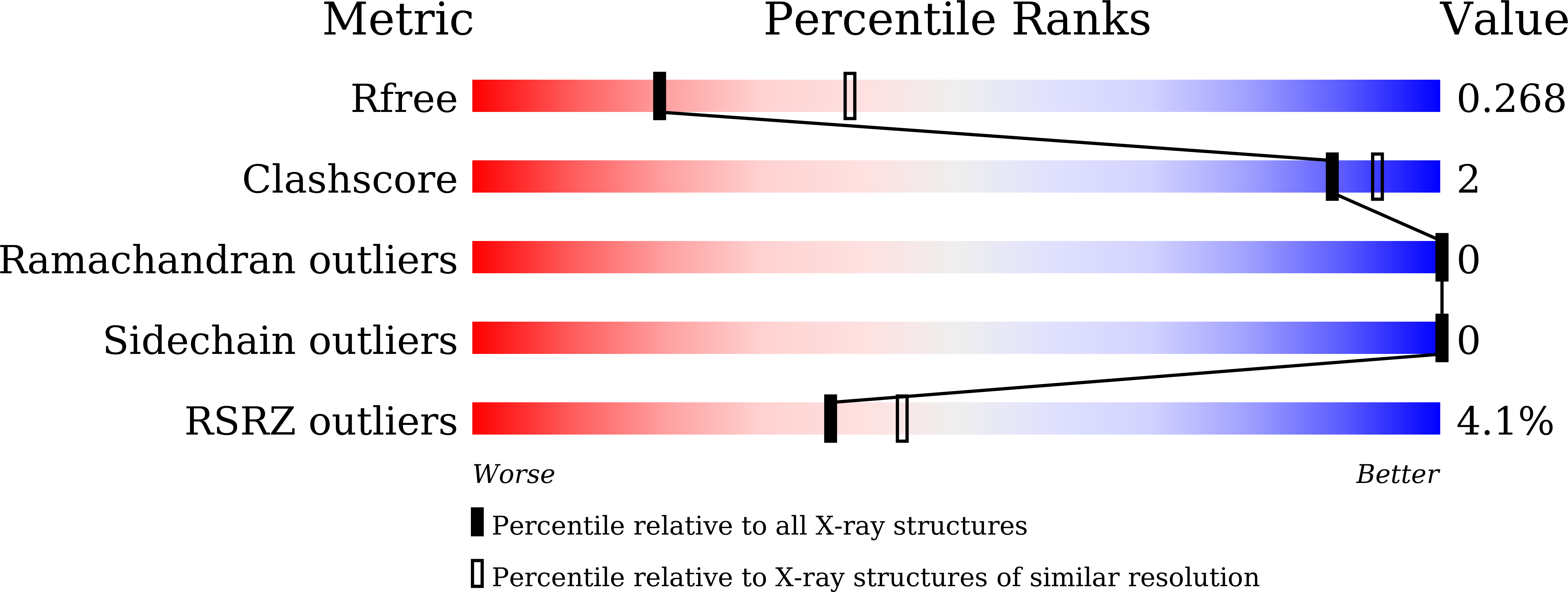
Resolution:
2.76 Å
R-Value Free:
0.26
R-Value Work:
0.21
R-Value Observed:
0.21
Space Group:
P 21 21 21
Macromolecular Entities
Structures with similar UniProt ID
Protein Blast
Polymer Type:polypeptide(L)
Molecule:SIRPA protein
Chain IDs:A, B
Chain Length:142
Number of Molecules:2
Biological Source:Homo sapiens
Structures with similar UniProt ID
Protein Blast
Polymer Type:polypeptide(L)
Molecule:Leukocyte surface antigen CD47
Gene (Uniprot):CD47
Chain IDs:C, D
Chain Length:125
Number of Molecules:2
Biological Source:Homo sapiens
Modified Residue
Compound ID Chain ID Parent Comp ID Details 2D Image
PCA C GLN modified residue
Primary Citation
Crystal Structure of Human CD47 in Complex with Engineered SIRP alpha.D1(N80A).
Molecules 27 ? ? (2022)
PMID: 36080360 DOI: 10.3390/molecules27175574

Abstact

Background: Targeting the CD47/SIRPα signaling pathway represents a novel approach to enhance anti-tumor immunity. However, the crystal structure of the CD47/SIRPα has not been fully studied. This study aims to analyze the structure interface of the complex of CD47 and IMM01, a novel recombinant SIRPα-Fc fusion protein. Methods: IMM01-Fab/CD47 complex was crystalized, and diffraction images were collected. The complex structure was determined by molecular replacement using the program PHASER with the CD47-SIRPαv2 structure (PDB code 2JJT) as a search model. The model was manually built using the COOT program and refined using TLS parameters in REFMAC from the CCP4 program suite. Results: Crystallization and structure determination analysis of the interface of IMM01/CD47 structure demonstrated CD47 surface buried by IMM01. Comparison with the literature structure (PDB ID 2JJT) showed that the interactions of IMM01/CD47 structure are the same. All the hydrogen bonds that appear in the literature structure are also present in the IMM01/CD47 structure. These common hydrogen bonds are stable under different crystal packing styles, suggesting that these hydrogen bonds are important for protein binding. In the structure of human CD47 in complex with human SIRPα, except SER66, the amino acids that form hydrogen bonds are all conserved. Furthermore, comparing with the structure of PDB ID 2JJT, the salt bridge interaction from IMM01/CD47 structure are very similar, except the salt bridge bond between LYS53 in IMM01 and GLU106 in CD47, which only occurs between the B and D chains. However, as the side chain conformation of LYS53 in chain A is slightly different, the salt bridge bond is absent between the A and C chains. At this site between chain A and chain C, there are a salt bridge bond between LYS53 (A) and GLU104 (C) and a salt bridge bond between HIS56 (A) and GLU106 (C) instead. According to the sequence alignment results of SIRPα, SIRPβ and SIRPγ in the literature of PDB ID 2JJT, except ASP100, the amino acids that form common salt bridge bonds are all conserved. Conclusion: Our data demonstrated crystal structure of the IMM01/CD47 complex and provides a structural basis for the structural binding interface and future clinical applications.

Legend

Protein

Chemical

Disease

Primary Citation of related structures