
Deposition Date
2022-07-01
Release Date
2023-11-22
Last Version Date
2024-03-13
Entry Detail
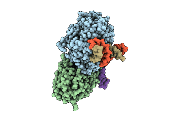
PDB ID:
7YCP
Keywords:
Title:
TR-SFX MmCPDII-DNA complex: 250 ps snapshot. Includes 250 ps, dark, and extrapolated structure factors
Biological Source:
Source Organism(s):
Methanosarcina mazei (Taxon ID: 192952)
synthetic construct (Taxon ID: 32630)
synthetic construct (Taxon ID: 32630)
Expression System(s):
Method Details:
Experimental Method:
Resolution:
2.08 Å
R-Value Free:
0.23
R-Value Work:
0.22
R-Value Observed:
0.22
Space Group:
P 21 21 21
