7Y9B image
Entry Detail
PDB ID:
7Y9B
Title:
Crystal structure of the membrane (M) protein of a SARS-COV-2-related coronavirus
Biological Source:
PDB Version:
Deposition Date:
2022-06-24
Release Date:
2022-08-17
Method Details:
Experimental Method:
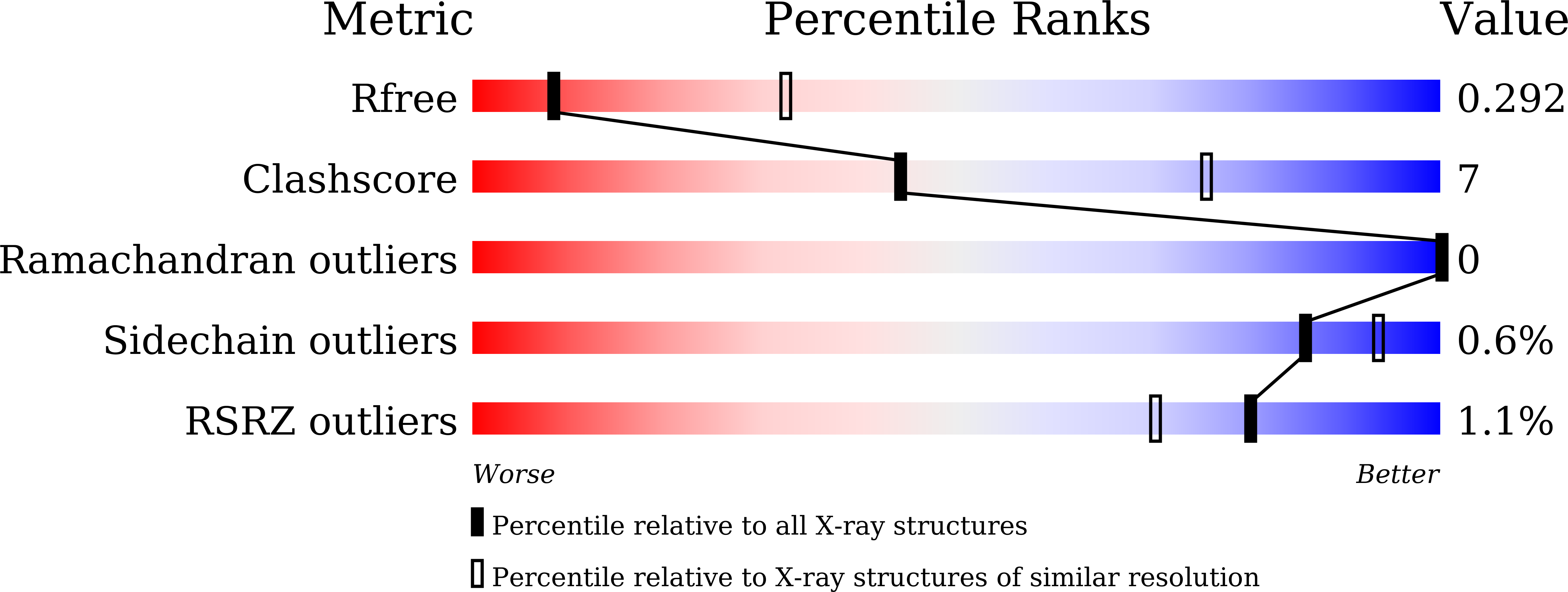
Resolution:
3.21 Å
R-Value Free:
0.29
R-Value Work:
0.27
R-Value Observed:
0.27
Space Group:
P 1 21 1
Macromolecular Entities
Polymer Type:polypeptide(L)
Description:Membrane protein
Chain IDs:A, B, C, D
Chain Length:220
Number of Molecules:4
Biological Source:Pipistrellus bat coronavirus HKU5
Ligand Molecules
Primary Citation
Crystal structure of the membrane (M) protein from a bat betacoronavirus.
Pnas Nexus 2 pgad021 pgad021 (2023)
PMID: 36874273 DOI: 10.1093/pnasnexus/pgad021

Abstact

The membrane (M) protein is the most abundant structural protein of coronaviruses including MERS-CoV, SARS-CoV, and SARS-CoV-2, and plays a central role in virus assembly through its interaction with various partner proteins. However, mechanistic details about how M protein interacts with others remain elusive due to lack of high-resolution structures. Here, we present the first crystal structure of a betacoronavirus M protein from Pipistrellus bat coronavirus HKU5 (batCOV5-M), which is closely related to MERS-CoV, SARS-CoV, and SARS-CoV-2 M proteins. Furthermore, an interaction analysis indicates that the carboxy-terminus of the batCOV5 nucleocapsid (N) protein mediates its interaction with batCOV5-M. Combined with a computational docking analysis an M-N interaction model is proposed, providing insight into the mechanism of M protein-mediated protein interactions.

Legend

Protein

Chemical

Disease

Primary Citation of related structures