Deposition Date
2022-06-22
Release Date
2023-04-26
Last Version Date
2024-11-20
Entry Detail
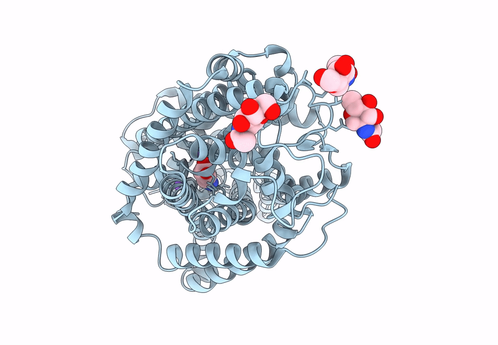
PDB ID:
7Y7W
Keywords:
Title:
Cryo-EM structure of human GABA transporter GAT1 bound with GABA in NaCl solution in an inward-occluded state at 2.4 angstrom
Biological Source:
Source Organism(s):
Homo sapiens (Taxon ID: 9606)
Expression System(s):
Method Details:
Experimental Method:
Resolution:
2.40 Å
Aggregation State:
PARTICLE
Reconstruction Method:
SINGLE PARTICLE