
Deposition Date
2022-06-08
Release Date
2023-06-14
Last Version Date
2023-10-11
Entry Detail
PDB ID:
7Y1K
Keywords:
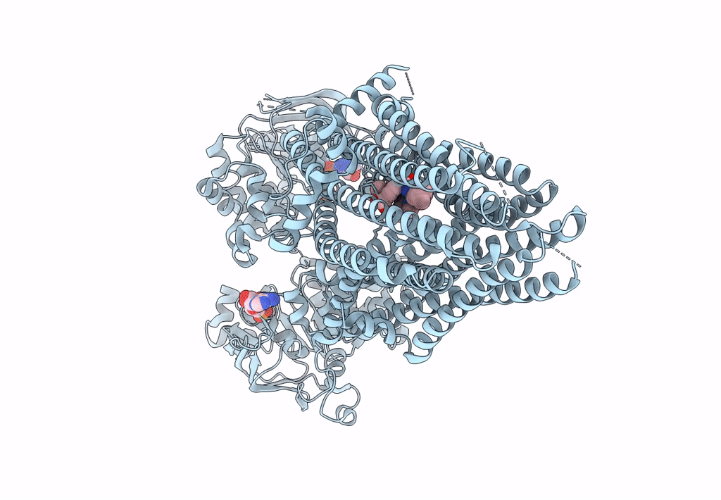
Title:
Structure of SUR2A in complex with Mg-ATP, Mg-ADP and repaglinide in the inward-facing conformation
Biological Source:
Source Organism(s):
Rattus norvegicus (Taxon ID: 10116)
Expression System(s):
Method Details:
Experimental Method:
Resolution:
3.80 Å
Aggregation State:
PARTICLE
Reconstruction Method:
SINGLE PARTICLE
