Deposition Date
2022-06-03
Release Date
2022-12-21
Last Version Date
2023-11-29
Entry Detail
PDB ID:
7Y02
Keywords:
Title:
Crystal structure of Ricin A chain bound with (S)-2-(2-amino-4-oxo-3,4-dihydropteridine-7-carboxamido)-3-(4-fluorophenyl)propanoic acid
Biological Source:
Source Organism(s):
Ricinus communis (Taxon ID: 3988)
Expression System(s):
Method Details:
Experimental Method:
Resolution:
1.60 Å
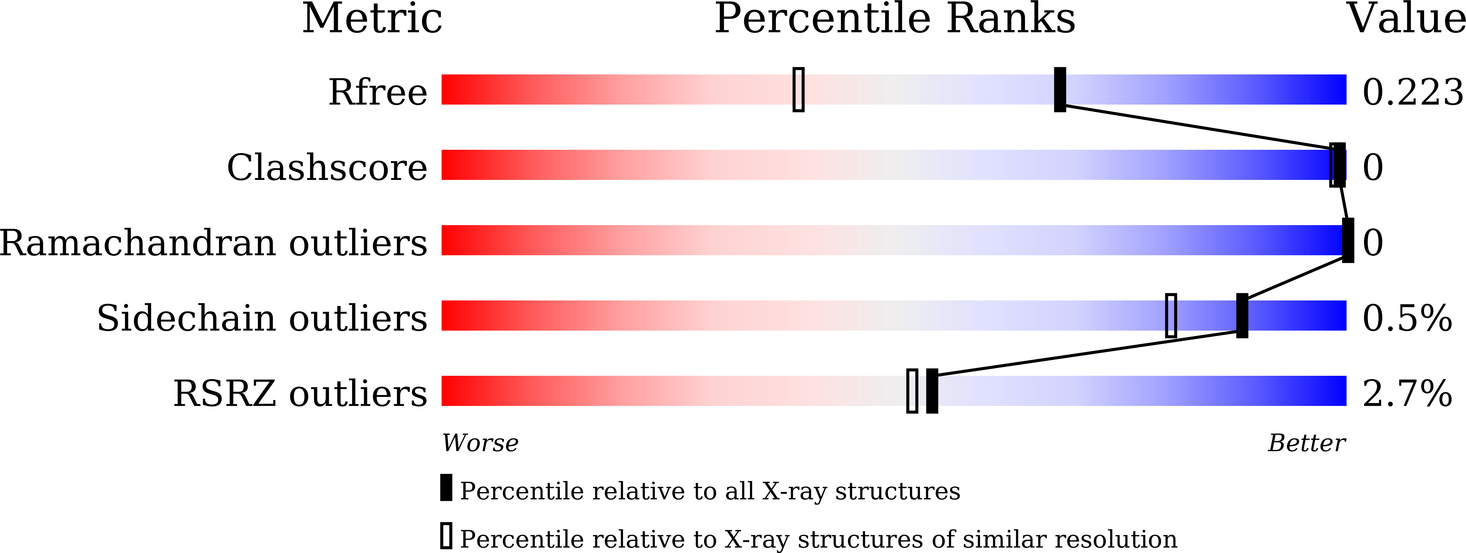
R-Value Free:
0.22
R-Value Work:
0.20
R-Value Observed:
0.20
Space Group:
P 41 21 2