Deposition Date
2022-06-02
Release Date
2023-07-12
Last Version Date
2023-11-29
Entry Detail
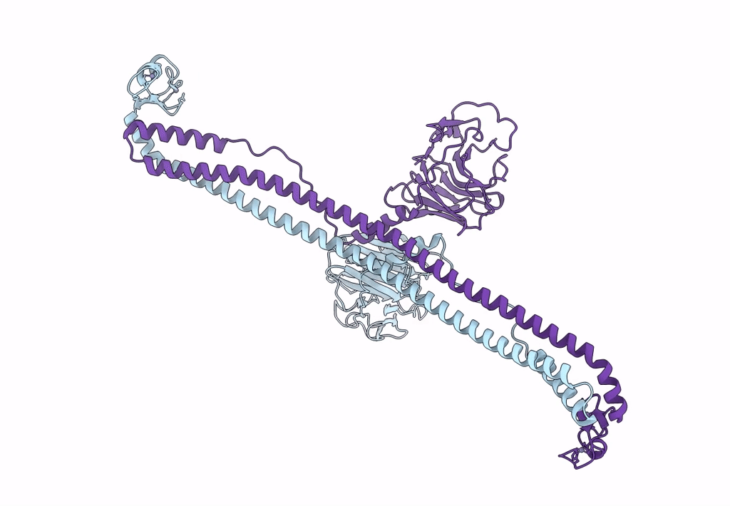
Biological Source:
Source Organism(s):
Mus musculus (Taxon ID: 10090)
Expression System(s):
Method Details:
Experimental Method:
Resolution:
3.28 Å
R-Value Free:
0.30
R-Value Work:
0.25
R-Value Observed:
0.25
Space Group:
C 1 2 1