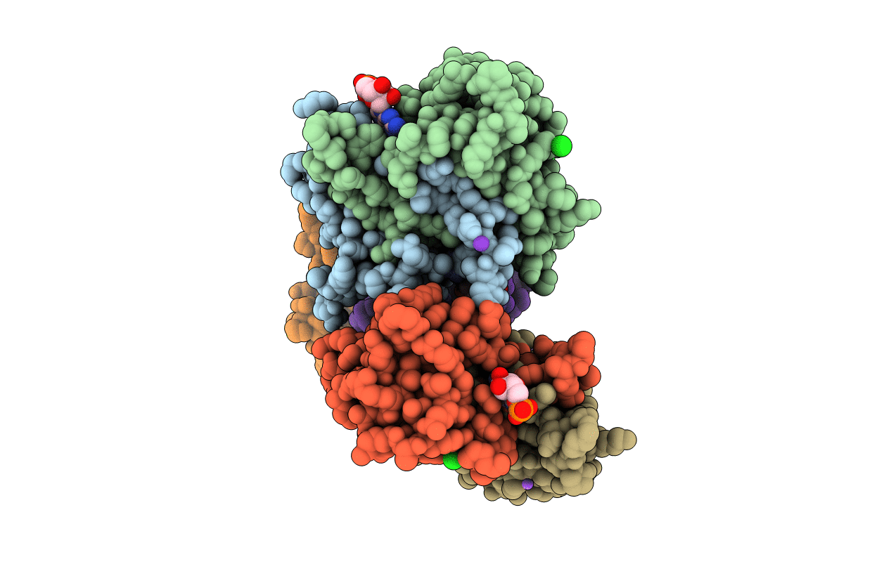
7XXK image
Entry Detail
PDB ID:
7XXK
Keywords:
Title:
Crystal structure of SARS-CoV-2 N-CTD in complex with GMP
Biological Source:
PDB Version:
Deposition Date:
2022-05-30
Release Date:
2022-06-29
Method Details:
Experimental Method:
Resolution:
2.00 Å
R-Value Free:
0.23
R-Value Work:
0.17
R-Value Observed:
0.18
Space Group:
P 21 21 21
Macromolecular Entities
Polymer Type:polypeptide(L)
Description:Nucleoprotein
Chain IDs:A, B, C, D, E, F
Chain Length:120
Number of Molecules:6
Biological Source:Severe acute respiratory syndrome coronavirus 2
Primary Citation
Structural basis of the C-terminal domain of SARS-CoV-2 N protein in complex with GMP reveals critical residues for RNA interaction.
Bioorg.Med.Chem.Lett. ? 130014 130014 (2024)
PMID: 39489230 DOI: 10.1016/j.bmcl.2024.130014

Abstact

The severe acute respiratory syndrome coronavirus 2 (SARS-CoV-2) nucleocapsid (N) protein performs multiple functions during the viral life cycle, particularly in binding to the viral genomic RNA to form a helical ribonucleoprotein complex. Here, we present that the C-terminal domain of SARS-CoV-2 N protein (N-CTD) specifically interacts with polyguanylic acid (poly(G)). The crystal structure of the N-CTD in complex with 5'-guanylic acid (GMP, also known as guanosine monophosphate) was determined at a resolution of approximately 2.0 Å. A novel GMP-binding pocket in the N-CTD was illustrated. Residues Arg259 and Lys338 were identified to play key roles in binding to GMP through mutational analysis. These two residues are absolutely conserved in the other two highly pathogenic CoVs, SARS-CoV and Middle East respiratory syndrome coronavirus (MERS-CoV). Overall, our findings expand the structural information on N protein interacting with guanylate and reveal a conserved GMP-binding pocket as a potential antiviral target.

Legend

Protein

Chemical

Disease

Primary Citation of related structures