Planned Maintenance: Some services may turn out to be unavailable from 15th January, 2026 to 16th January, 2026. We apologize for the inconvenience!

7XWM image
Deposition Date 2022-05-26
Release Date 2023-04-12
Last Version Date 2023-11-29
Entry Detail
PDB ID:
7XWM
Keywords:
Title:
structure of patulin-detoxifying enzyme Y155F/V187K with NADPH
Biological Source:
Source Organism(s):
Expression System(s):
Method Details:
Experimental Method:
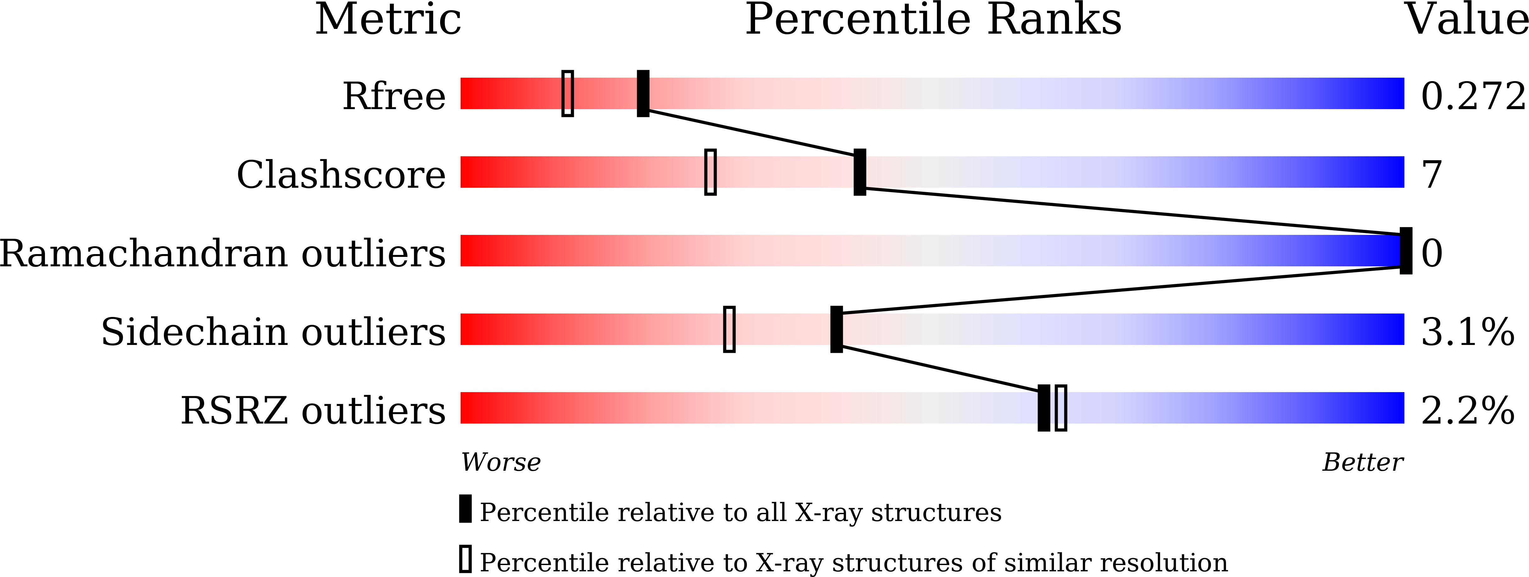
Resolution:
1.98 Å
R-Value Free:
0.26
R-Value Work:
0.19
R-Value Observed:
0.20
Space Group:
P 1 21 1
Macromolecular Entities
Structures with similar UniProt ID
Protein Blast
Polymer Type:polypeptide(L)
Molecule:Short-chain dehydrogenase/reductase
Gene (Uniprot):SDR
Mutagens:Y155F, V187F
Chain IDs:A, B, C, D
Chain Length:261
Number of Molecules:4
Biological Source:Meyerozyma guilliermondii
Ligand Molecules
Primary Citation
Structure-based rational design of a short-chain dehydrogenase/reductase for improving activity toward mycotoxin patulin.
Int.J.Biol.Macromol. 222 421 428 (2022)
PMID: 36176222 DOI: 10.1016/j.ijbiomac.2022.09.121

Abstact

Patulin is a fatal mycotoxin that is widely detected in drinking water and fruit-derived products contaminated by diverse filamentous fungi. CgSDR from Candida guilliermondii represents the first NADPH-dependent short-chain dehydrogenase/reductase that catalyzes the reduction of patulin to the nontoxic E-ascladiol. To elucidate the catalytic mechanism of CgSDR, we solved its crystal structure in complex with cofactor and substrate. Structural analyses indicate that patulin is situated in a hydrophobic pocket adjacent to the cofactor, with the hemiacetal ring orienting toward the nicotinamide moiety of NADPH. In addition, we conducted structure-guided engineering to modify substrate-binding residue V187 and obtained variant V187F, V187K and V187W, whose catalytic activity was elevated by 3.9-, 2.2- and 1.7-fold, respectively. The crystal structures of CgSDR variants suggest that introducing additional aromatic stacking or hydrogen-bonding interactions to bind the lactone ring of patulin might account for the observed enhanced activity. These results illustrate the catalytic mechanism of SDR-mediated patulin detoxification for the first time and provide the upgraded variants that exhibit tremendous potentials in industrial applications.

Legend

Protein

Chemical

Disease

Primary Citation of related structures
Feedback Form
Name
Email
Institute
Feedback