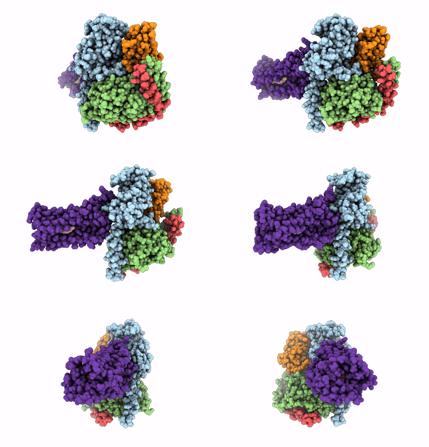
7XV3 image
Deposition Date 2022-05-20
Release Date 2023-02-15
Last Version Date 2024-11-13
Entry Detail
PDB ID:
7XV3
Title:
Cryo-EM structure of LPS-bound GPR174 in complex with Gs protein
Biological Source:
Source Organism:
Homo sapiens (Taxon ID: 9606)
Lama glama (Taxon ID: 9844)
Host Organism:
Method Details:
Experimental Method:
Resolution:
2.76 Å
Aggregation State:
PARTICLE
Reconstruction Method:
SINGLE PARTICLE
Macromolecular Entities
Polymer Type:polypeptide(L)
Molecule:Engineered G protein subunit S (mini-Gs)
Chain IDs:C (auth: A)
Chain Length:361
Number of Molecules:1
Biological Source:Homo sapiens
Structures with similar UniProt ID
Protein Blast
Polymer Type:polypeptide(L)
Molecule:Guanine nucleotide-binding protein G(I)/G(S)/G(T) subunit beta-1
Gene (Uniprot):GNB1
Chain IDs:E (auth: B)
Chain Length:345
Number of Molecules:1
Biological Source:Homo sapiens
Structures with similar UniProt ID
Protein Blast
Polymer Type:polypeptide(L)
Molecule:Guanine nucleotide-binding protein G(I)/G(S)/G(O) subunit gamma-2
Gene (Uniprot):GNG2
Chain IDs:B (auth: G)
Chain Length:71
Number of Molecules:1
Biological Source:Homo sapiens
Polymer Type:polypeptide(L)
Molecule:Nb35
Chain IDs:D (auth: N)
Chain Length:161
Number of Molecules:1
Biological Source:Lama glama
Structures with similar UniProt ID
Protein Blast
Polymer Type:polypeptide(L)
Molecule:Probable G-protein coupled receptor 174
Gene (Uniprot):GPR174
Chain IDs:A (auth: R)
Chain Length:333
Number of Molecules:1
Biological Source:Homo sapiens
Ligand Molecules
Primary Citation
Structural basis of lysophosphatidylserine receptor GPR174 ligand recognition and activation.
Nat Commun 14 1012 1012 (2023)
PMID: 36823105 DOI: 10.1038/s41467-023-36575-0

Abstact

Lysophosphatidylserine (LysoPS) is a lipid mediator that induces multiple cellular responses through binding to GPR174. Here, we present the cryo-electron microscopy (cryo-EM) structure of LysoPS-bound human GPR174 in complex with Gs protein. The structure reveals a ligand recognition mode, including the negatively charged head group of LysoPS forms extensive polar interactions with surrounding key residues of the ligand binding pocket, and the L-serine moiety buries deeply into a positive charged cavity in the pocket. In addition, the structure unveils a partially open pocket on transmembrane domain helix (TM) 4 and 5 for a lateral entry of ligand. Finally, the structure reveals a Gs engaging mode featured by a deep insertion of a helix 5 (αH5) and extensive polar interactions between receptor and αH5. Taken together, the information revealed by our structural study provides a framework for understanding LysoPS signaling and a rational basis for designing LysoPS receptor-targeting drugs.

Legend

Protein

Chemical

Disease

Primary Citation of related structures