7XK3 image
Entry Detail
PDB ID:
7XK3
EMDB ID:
Keywords:
Title:
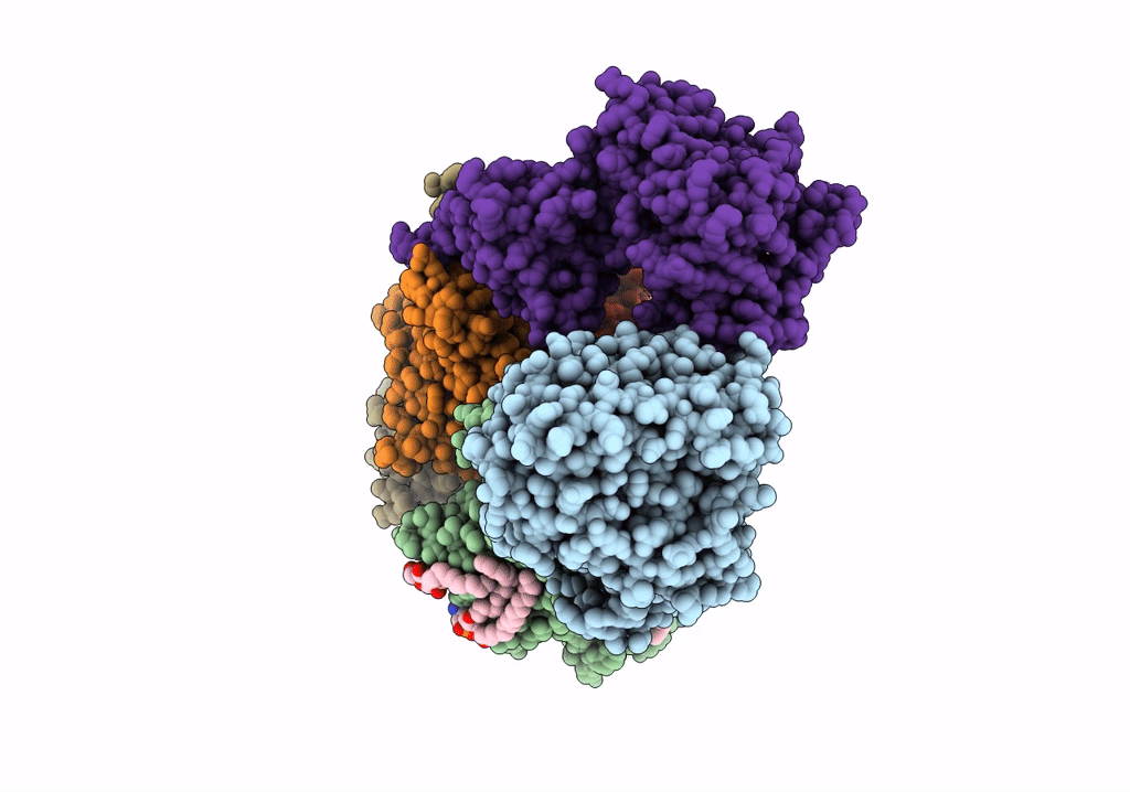
Cryo-EM structure of Na+-pumping NADH-ubiquinone oxidoreductase from Vibrio cholerae, state 1
Biological Source:
Source Organism:
Host Organism:
PDB Version:
Deposition Date:
2022-04-19
Release Date:
2022-07-20
Method Details:
Experimental Method:
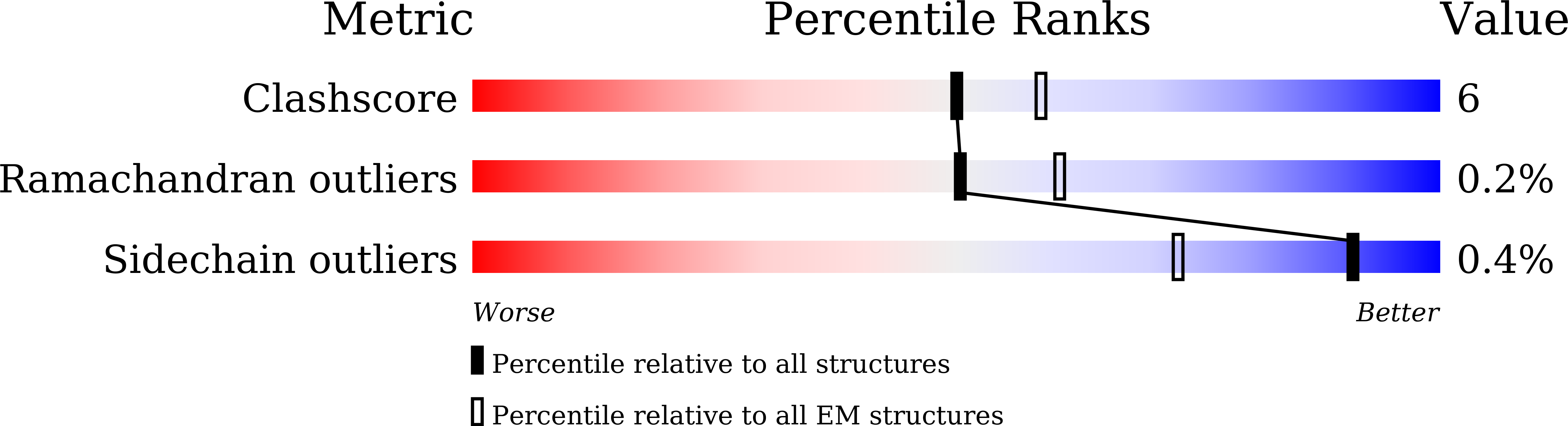
Resolution:
3.10 Å
Aggregation State:
PARTICLE
Reconstruction Method:
SINGLE PARTICLE
Macromolecular Entities
Polymer Type:polypeptide(L)
Description:Na(+)-translocating NADH-quinone reductase subunit A
Chain IDs:A
Chain Length:446
Number of Molecules:1
Biological Source:Vibrio cholerae O395
Polymer Type:polypeptide(L)
Description:Na(+)-translocating NADH-quinone reductase subunit B
Chain IDs:B
Chain Length:415
Number of Molecules:1
Biological Source:Vibrio cholerae O395
Polymer Type:polypeptide(L)
Description:Na(+)-translocating NADH-quinone reductase subunit C
Chain IDs:C
Chain Length:257
Number of Molecules:1
Biological Source:Vibrio cholerae O395
Polymer Type:polypeptide(L)
Description:Na(+)-translocating NADH-quinone reductase subunit D
Chain IDs:D
Chain Length:210
Number of Molecules:1
Biological Source:Vibrio cholerae O395
Polymer Type:polypeptide(L)
Description:Na(+)-translocating NADH-quinone reductase subunit E
Chain IDs:E
Chain Length:198
Number of Molecules:1
Biological Source:Vibrio cholerae O395
Polymer Type:polypeptide(L)
Description:Na(+)-translocating NADH-quinone reductase subunit F
Chain IDs:F
Chain Length:414
Number of Molecules:1
Biological Source:Vibrio cholerae O395
Primary Citation
Cryo-EM structures of Na + -pumping NADH-ubiquinone oxidoreductase from Vibrio cholerae.
Nat Commun 13 4082 4082 (2022)
PMID: 35882843 DOI: 10.1038/s41467-022-31718-1

Abstact

The Na+-pumping NADH-ubiquinone oxidoreductase (Na+-NQR) couples electron transfer from NADH to ubiquinone with Na+-pumping, generating an electrochemical Na+ gradient that is essential for energy-consuming reactions in bacteria. Since Na+-NQR is exclusively found in prokaryotes, it is a promising target for highly selective antibiotics. However, the molecular mechanism of inhibition is not well-understood for lack of the atomic structural information about an inhibitor-bound state. Here we present cryo-electron microscopy structures of Na+-NQR from Vibrio cholerae with or without a bound inhibitor at 2.5- to 3.1-Å resolution. The structures reveal the arrangement of all six redox cofactors including a herein identified 2Fe-2S cluster located between the NqrD and NqrE subunits. A large part of the hydrophilic NqrF is barely visible in the density map, suggesting a high degree of flexibility. This flexibility may be responsible to reducing the long distance between the 2Fe-2S centers in NqrF and NqrD/E. Two different types of specific inhibitors bind to the N-terminal region of NqrB, which is disordered in the absence of inhibitors. The present study provides a foundation for understanding the function of Na+-NQR and the binding manner of specific inhibitors.

Legend

Protein

Chemical

Disease

Primary Citation of related structures