7XJD image
Deposition Date 2022-04-16
Release Date 2023-03-22
Last Version Date 2024-11-20
Entry Detail
PDB ID:
7XJD
Title:
Crystal structure of bacteriorhodopsin in the ground state by red laser irradiation
Biological Source:
Source Organism:
Method Details:
Experimental Method:
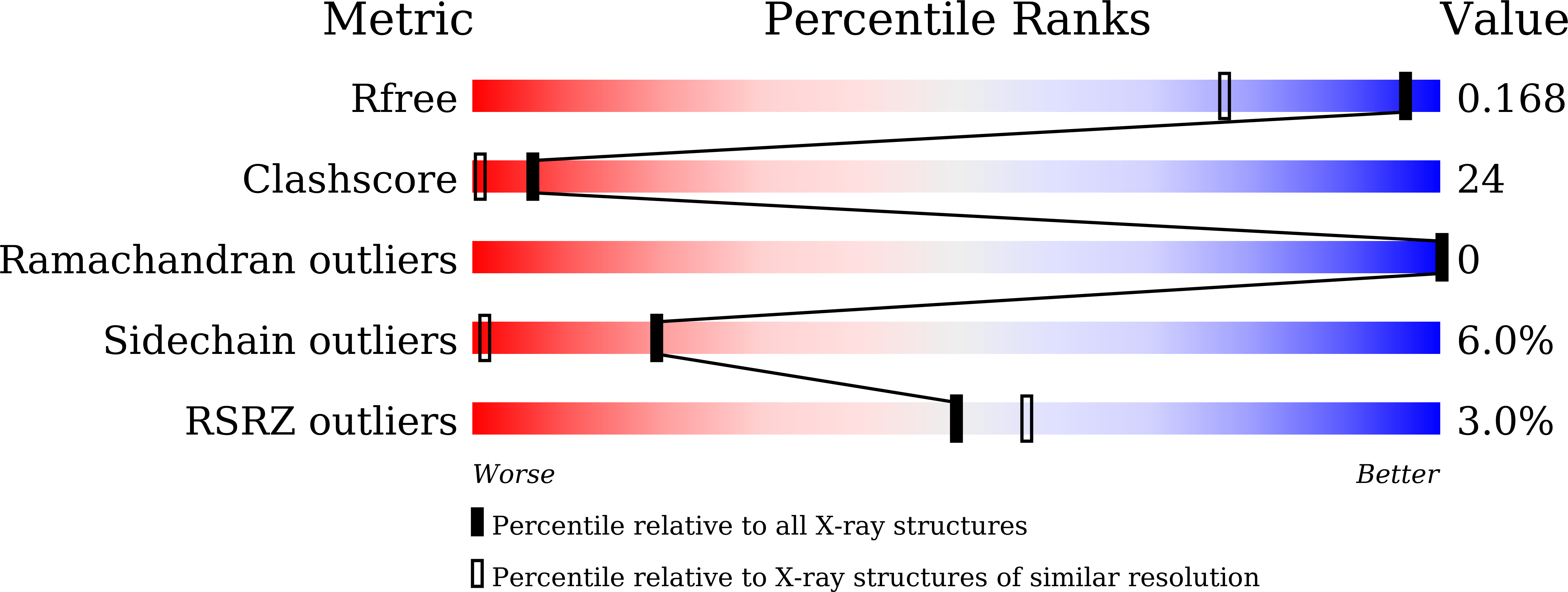
Resolution:
1.33 Å
R-Value Free:
0.17
R-Value Work:
0.13
Space Group:
P 63
Macromolecular Entities
Structures with similar UniProt ID
Protein Blast
Polymer Type:polypeptide(L)
Molecule:Bacteriorhodopsin
Gene (Uniprot):bop
Chain IDs:A
Chain Length:230
Number of Molecules:1
Biological Source:Halobacterium salinarum NRC-1
Primary Citation
Detailed analysis of distorted retinal and its interaction with surrounding residues in the K intermediate of bacteriorhodopsin.
Commun Biol 6 190 190 (2023)
PMID: 36808185 DOI: 10.1038/s42003-023-04554-2

Abstact

The K intermediate of proton pumping bacteriorhodopsin is the first intermediate generated after isomerization of retinal to the 13-cis form. Although various structures have been reported for the K intermediate until now, these differ from each other, especially in terms of the conformation of the retinal chromophore and its interaction with surrounding residues. We report here an accurate X-ray crystallographic analysis of the K structure. The polyene chain of 13-cis retinal is observed to be S-shaped. The side chain of Lys216, which is covalently bound to retinal via the Schiff-base linkage, interacts with residues, Asp85 and Thr89. In addition, the Nζ-H of the protonated Schiff-base linkage interacts with a residue, Asp212 and a water molecule, W402. Based on quantum chemical calculations for this K structure, we examine the stabilizing factors of distorted conformation of retinal and propose a relaxation manner to the next L intermediate.

Legend

Protein

Chemical

Disease

Primary Citation of related structures
Feedback Form
Name
Email
Institute
Feedback