7XFW image
Deposition Date 2022-04-02
Release Date 2022-05-11
Last Version Date 2024-11-13
Entry Detail
PDB ID:
7XFW
Keywords:
Title:
Crystal structure of the ternary complex of Peptidoglycan recognition protein, PGRP-S with hexanoic and tartaric acids at 2.07 A resolution.
Biological Source:
Source Organism:
Method Details:
Experimental Method:
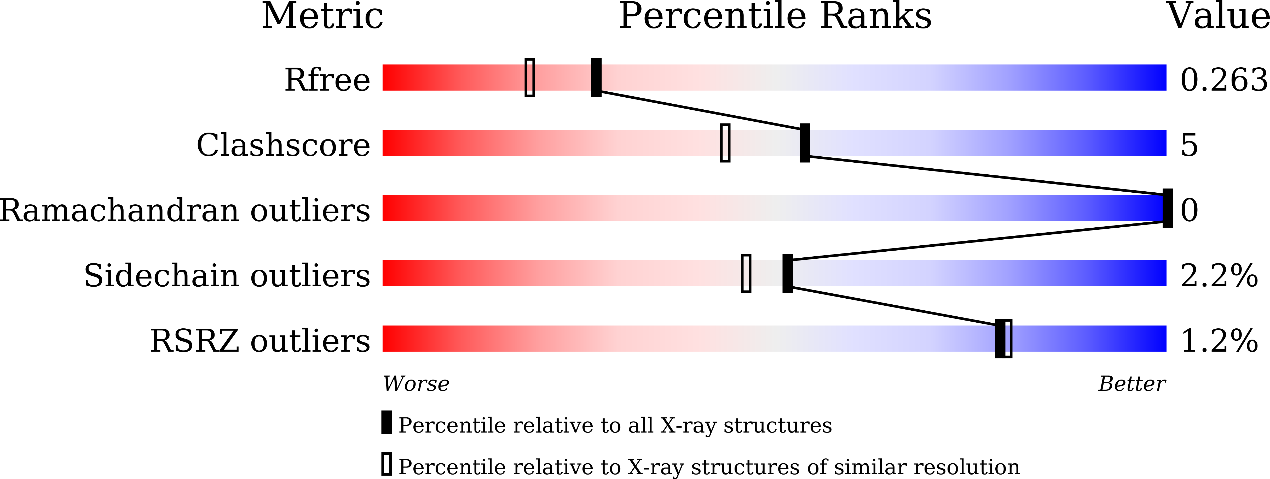
Resolution:
2.07 Å
R-Value Free:
0.25
R-Value Work:
0.19
Space Group:
I 2 2 2
Macromolecular Entities
Structures with similar UniProt ID
Protein Blast
Polymer Type:polypeptide(L)
Molecule:Peptidoglycan recognition protein 1
Gene (Uniprot):PGLYRP1
Chain IDs:A, B, C, D
Chain Length:171
Number of Molecules:4
Biological Source:Camelus dromedarius
Primary Citation
Structure of the complex of camel peptidoglycan recognition protein-S with hexanoic acid reveals novel features of the versatile ligand-binding site at the dimeric interface.
Biochim Biophys Acta Proteins Proteom 1871 140887 140887 (2022)
PMID: 36596432 DOI: 10.1016/j.bbapap.2022.140887

Abstact

The short peptidoglycan recognition protein (PGRP-S) of the innate immune system recognizes the invading microbes through binding to their cell wall molecules. In order to understand the mode of binding of PGRP-S to bacterial cell wall molecules, the structure of the complex of camel PGRP-S (CPGRP-S) with hexanoic acid has been determined at 2.07 Å resolution. Previously, we had reported the structures of CPGRP-S in the native unbound state as well as in the complexed forms with the components of various bacterial cell wall molecules such as peptidoglycan (PGN), lipopolysaccharide (LPS), lipoteichoic acid (LTA), mycolic acid (MA) and other fatty acids. These structures revealed that CPGRP-S formed two homodimers which were designated as A-B and CD dimers. It also showed that the fatty acids bind to CPGRP-S in the binding site at the A-B dimer while the non-fatty acids were shown to bind at the interfaces of both A-B and CD dimers. The present structure of the complex of CPGRP-S with hexanoic acid (HA) showed that HA binds to CPGRP-S at the interface of CD dimer. HA was located in the same groove at the CD interface which was occupied by non-fatty acids such as PGN, LPS and LTA and interacts with residues from both C and D molecules. HA is firmly held in the groove with several hydrogen bonds and a number of van der Waals contacts. This is the first structure which reports the binding of a fatty acid in the cleft at the interface of CD dimer.

Legend

Protein

Chemical

Disease

Primary Citation of related structures