7XEB image
Entry Detail
PDB ID:
7XEB
Keywords:
Title:
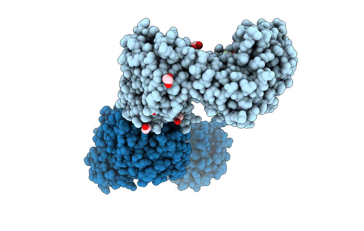
Collagenase from Grimontia (Vibrio) hollisae 1706B complexed with Gly-Pro-Hyp
Biological Source:
Host Organism:
PDB Version:
Deposition Date:
2022-03-30
Release Date:
2022-06-29
Method Details:
Experimental Method:
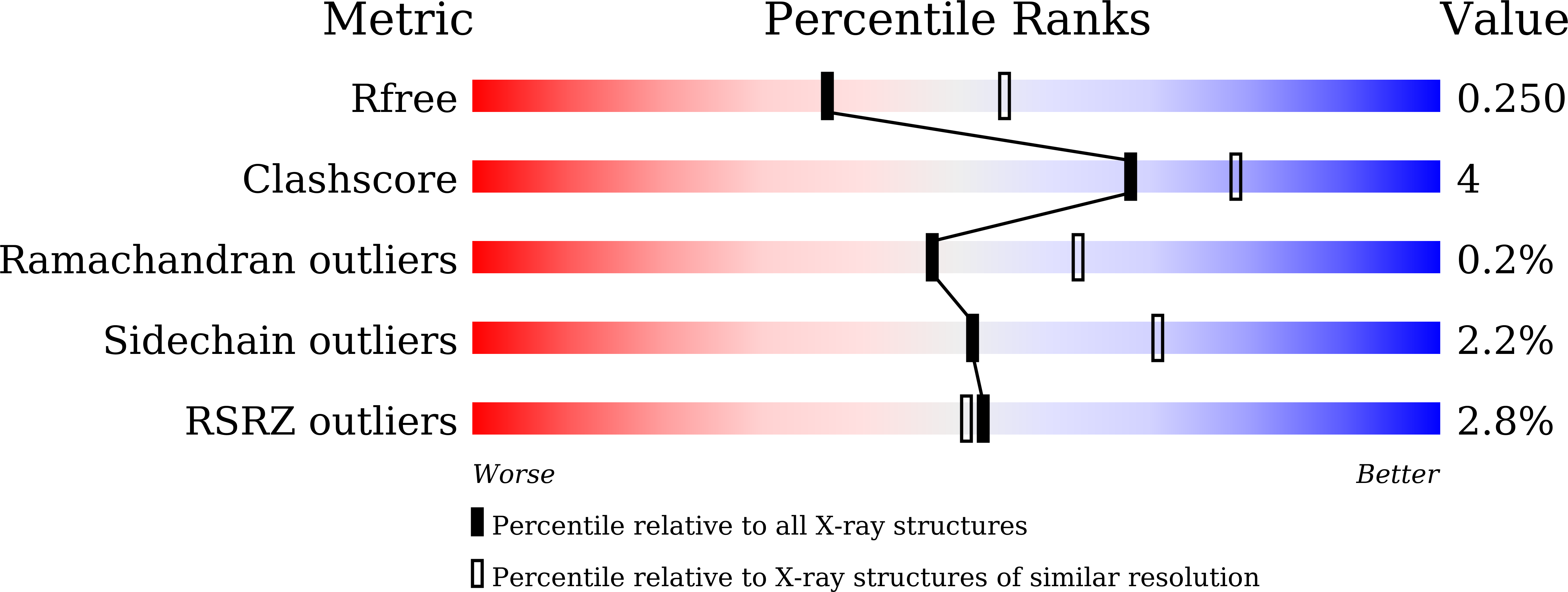
Resolution:
2.39 Å
R-Value Free:
0.25
R-Value Work:
0.20
R-Value Observed:
0.20
Space Group:
P 1 21 1
Macromolecular Entities
Polymer Type:polypeptide(L)
Description:Microbial collagenase
Chain IDs:A, B
Chain Length:559
Number of Molecules:2
Biological Source:Grimontia hollisae
Polymer Type:polypeptide(L)
Description:GLY-PRO-HYP peptide
Chain IDs:C, D, G, H
Chain Length:3
Number of Molecules:4
Biological Source:synthetic construct
Polymer Type:polypeptide(L)
Description:GLY-PRO-HYP-GLY-PRO-HYP peptide
Chain IDs:E, F, I, J
Chain Length:6
Number of Molecules:4
Biological Source:synthetic construct
Primary Citation
Crystal structure of Grimontia hollisae collagenase provides insights into its novel substrate specificity toward collagen.
J.Biol.Chem. 298 102109 102109 (2022)
PMID: 35679897 DOI: 10.1016/j.jbc.2022.102109

Abstact

Collagenase from the gram-negative bacterium Grimontia hollisae strain 1706B (Ghcol) degrades collagen more efficiently even than clostridial collagenase, the most widely used industrial collagenase. However, the structural determinants facilitating this efficiency are unclear. Here, we report the crystal structures of ligand-free and Gly-Pro-hydroxyproline (Hyp)-complexed Ghcol at 2.2 and 2.4 Å resolution, respectively. These structures revealed that the activator and peptidase domains in Ghcol form a saddle-shaped structure with one zinc ion and four calcium ions. In addition, the activator domain comprises two homologous subdomains, whereas zinc-bound water was observed in the ligand-free Ghcol. In the ligand-complexed Ghcol, we found two Gly-Pro-Hyp molecules, each bind at the active site and at two surfaces on the duplicate subdomains of the activator domain facing the active site, and the nucleophilic water is replaced by the carboxyl oxygen of Hyp at the P1 position. Furthermore, all Gly-Pro-Hyp molecules bound to Ghcol have almost the same conformation as Pro-Pro-Gly motif in model collagen (Pro-Pro-Gly)10, suggesting these three sites contribute to the unwinding of the collagen triple helix. A comparison of activities revealed that Ghcol exhibits broader substrate specificity than clostridial collagenase at the P2 and P2' positions, which may be attributed to the larger space available for substrate binding at the S2 and S2' sites in Ghcol. Analysis of variants of three active-site Tyr residues revealed that mutation of Tyr564 affected catalysis, whereas mutation of Tyr476 or Tyr555 affected substrate recognition. These results provide insights into the substrate specificity and mechanism of G. hollisae collagenase.

Legend

Protein

Chemical

Disease

Primary Citation of related structures