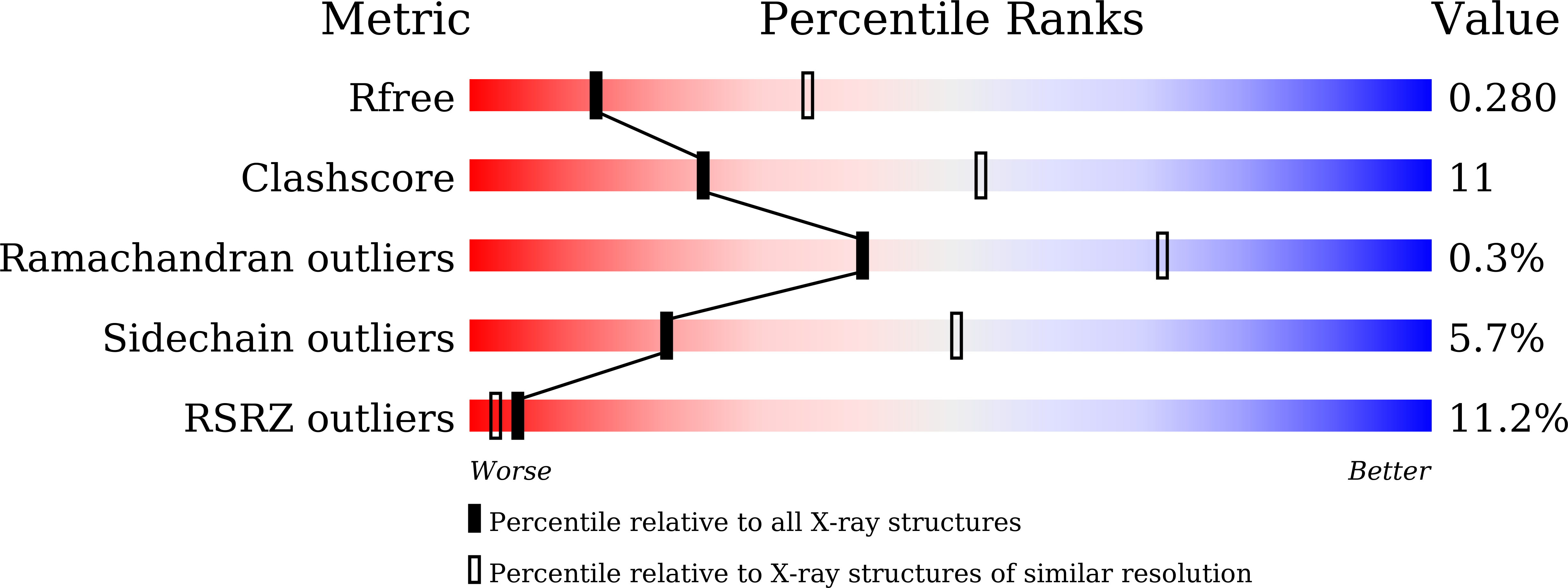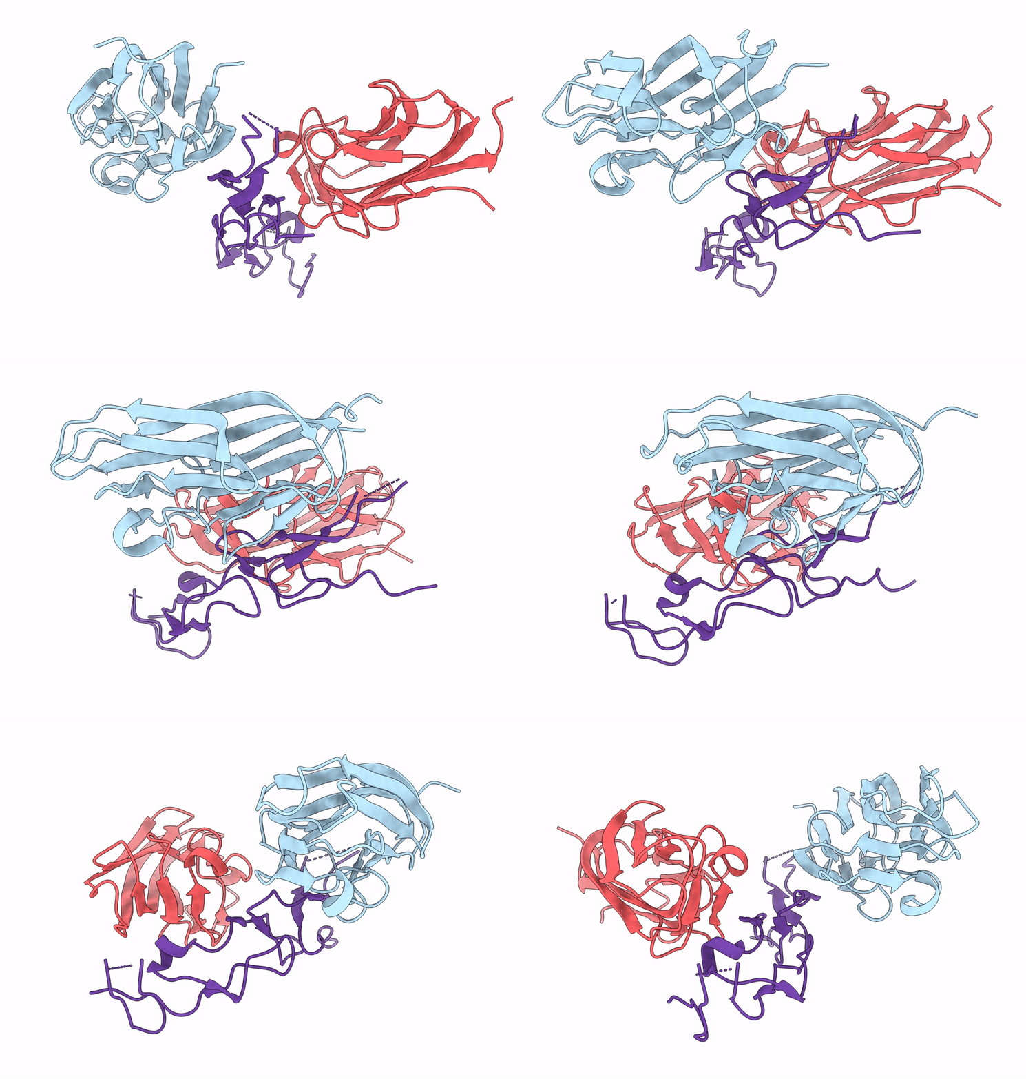
7X9G image
Deposition Date 2022-03-15
Release Date 2023-02-22
Last Version Date 2024-11-20
Entry Detail
PDB ID:
7X9G
Title:
Crystal structure of human EDA and EDAR
Biological Source:
Source Organism:
Homo sapiens (Taxon ID: 9606)
Method Details:
Experimental Method:
Resolution:
2.80 Å
R-Value Free:
0.28
R-Value Work:
0.24
R-Value Observed:
0.24
Space Group:
H 3
Macromolecular Entities
Polymer Type:polypeptide(L)
Molecule:Ectodysplasin-A, secreted form
Gene (Uniprot):EDA
Chain IDs:A, B
Chain Length:159
Number of Molecules:2
Biological Source:Homo sapiens
Polymer Type:polypeptide(L)
Molecule:Tumor necrosis factor receptor superfamily member EDAR
Gene (Uniprot):EDAR
Chain IDs:C
Chain Length:123
Number of Molecules:1
Biological Source:Homo sapiens
Ligand Molecules
Primary Citation
Structural insights into pathogenic mechanism of hypohidrotic ectodermal dysplasia caused by ectodysplasin A variants.
Nat Commun 14 767 767 (2023)
PMID: 36765055 DOI: 10.1038/s41467-023-36367-6

Abstact

EDA is a tumor necrosis factor (TNF) family member, which functions together with its cognate receptor EDAR during ectodermal organ development. Mutations of EDA have long been known to cause X-linked hypohidrotic dysplasia in humans characterized by primary defects in teeth, hair and sweat glands. However, the structural information of EDA interaction with EDAR is lacking and the pathogenic mechanism of EDA variants is poorly understood. Here, we report the crystal structure of EDA C-terminal TNF homology domain bound to the N-terminal cysteine-rich domains of EDAR. Together with biochemical, cellular and mouse genetic studies, we show that different EDA mutations lead to varying degrees of ectodermal developmental defects in mice, which is consistent with the clinical observations on human patients. Our work extends the understanding of the EDA signaling mechanism, and provides important insights into the molecular pathogenesis of disease-causing EDA variants.

Legend

Protein

Chemical

Disease

Primary Citation of related structures