Deposition Date
2022-03-10
Release Date
2022-09-07
Last Version Date
2025-08-06
Entry Detail
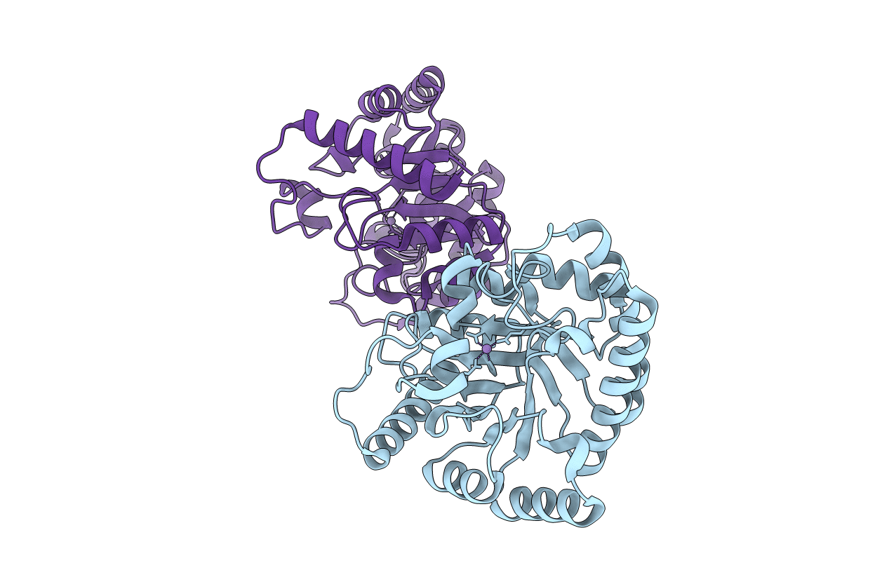
PDB ID:
7X7W
Keywords:
Title:
The X-ray Crystallographic Structure of D-Psicose 3-epimerase from Clostridia bacterium
Biological Source:
Source Organism(s):
Clostridia bacterium (Taxon ID: 2044939)
Expression System(s):
Method Details:
Experimental Method:
Resolution:
2.10 Å
R-Value Free:
0.25
R-Value Work:
0.20
R-Value Observed:
0.20
Space Group:
I 1 2 1