7X4O image
Deposition Date 2022-03-03
Release Date 2022-05-18
Last Version Date 2024-04-03
Entry Detail
PDB ID:
7X4O
Title:
Crystal structure of Vps17p PX from S. cerevisiae (Space)
Biological Source:
Source Organism:
Host Organism:
Method Details:
Experimental Method:
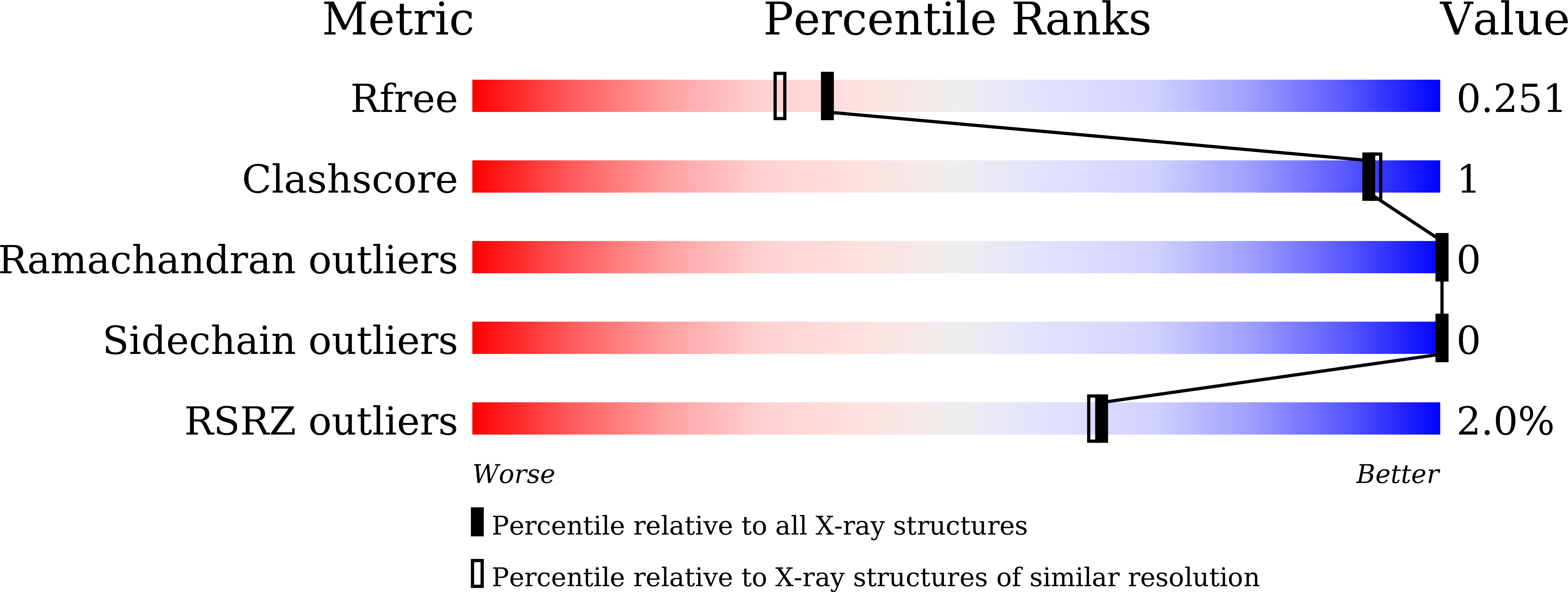
Resolution:
2.02 Å
R-Value Free:
0.25
R-Value Work:
0.19
R-Value Observed:
0.20
Space Group:
P 21 3
Macromolecular Entities
Structures with similar UniProt ID
Protein Blast
Polymer Type:polypeptide(L)
Molecule:Vacuolar protein sorting-associated protein 17
Gene (Uniprot):VPS17
Chain IDs:A, B
Chain Length:139
Number of Molecules:2
Biological Source:Saccharomyces cerevisiae S288C
Ligand Molecules
Primary Citation
Crystal structure of the PX domain of Vps17p from Saccharomyces cerevisiae.
Acta Crystallogr.,Sect.F 78 210 216 (2022)
PMID: 35506766 DOI: 10.1107/S2053230X22004472

Abstact

The structure determination of the PX (phox homology) domain of the Saccharomyces cerevisiae Vps17p protein presented a challenging case for molecular replacement because it has noncrystallographic symmetry close to a crystallographic axis. The combination of diffraction-quality crystals grown under microgravity on the International Space Station and a highly accurate template structure predicted by AlphaFold2 provided the key to successful crystal structure determination. Although the structure of the Vps17p PX domain is seen in many PX domains, no basic residues are found around the canonical phosphatidylinositol phosphate (PtdIns-P) binding site, suggesting an inability to bind PtdIns-P molecules.

Legend

Protein

Chemical

Disease

Primary Citation of related structures