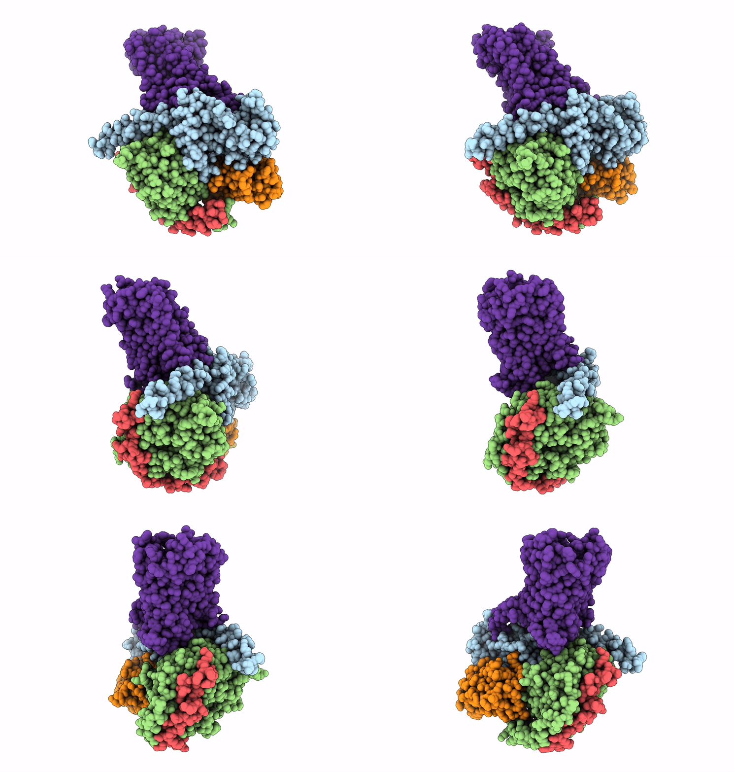
7WUQ image
Entry Detail
PDB ID:
7WUQ
EMDB ID:
Title:
Tethered peptide activation mechanism of adhesion GPCRs ADGRG2 and ADGRG4
Biological Source:
PDB Version:
Deposition Date:
2022-02-09
Release Date:
2022-04-27
Method Details:
Experimental Method:
Resolution:
2.90 Å
Aggregation State:
PARTICLE
Reconstruction Method:
SINGLE PARTICLE
Macromolecular Entities
Polymer Type:polypeptide(L)
Description:Guanine nucleotide-binding protein G(s) subunit alpha isoforms short
Mutations:S54N, G226A, E268A, N271K, K274D, R280K, T284D I285T
Chain IDs:A
Chain Length:394
Number of Molecules:1
Biological Source:Homo sapiens
Polymer Type:polypeptide(L)
Description:Guanine nucleotide-binding protein G(I)/G(S)/G(T) subunit beta-1
Chain IDs:B
Chain Length:358
Number of Molecules:1
Biological Source:Homo sapiens
Polymer Type:polypeptide(L)
Description:Guanine nucleotide-binding protein G(I)/G(S)/G(O) subunit gamma-2
Chain IDs:C (auth: G)
Chain Length:71
Number of Molecules:1
Biological Source:Homo sapiens
Polymer Type:polypeptide(L)
Description:Nanobody-35
Chain IDs:D (auth: N)
Chain Length:128
Number of Molecules:1
Biological Source:Lama glama
Polymer Type:polypeptide(L)
Description:Adhesion G-protein coupled receptor G2,mCherry
Chain IDs:E (auth: R)
Chain Length:683
Number of Molecules:1
Biological Source:Mus musculus
Ligand Molecules
Primary Citation
Tethered peptide activation mechanism of the adhesion GPCRs ADGRG2 and ADGRG4.
Nature 604 771 778 (2022)
PMID: 35418677 DOI: 10.1038/s41586-022-04590-8

Abstact

Adhesion G protein-coupled receptors (aGPCRs) constitute an evolutionarily ancient family of receptors that often undergo autoproteolysis to produce α and β subunits1-3. A tethered agonism mediated by the 'Stachel sequence' of the β subunit has been proposed to have central roles in aGPCR activation4-6. Here we present three cryo-electron microscopy structures of aGPCRs coupled to the Gs heterotrimer. Two of these aGPCRs are activated by tethered Stachel sequences-the ADGRG2-β-Gs complex and the ADGRG4-β-Gs complex (in which β indicates the β subunit of the aGPCR)-and the other is the full-length ADGRG2 in complex with the exogenous ADGRG2 Stachel-sequence-derived peptide agonist IP15 (ADGRG2(FL)-IP15-Gs). The Stachel sequences of both ADGRG2-β and ADGRG4-β assume a U shape and insert deeply into the seven-transmembrane bundles. Constituting the FXφφφXφ motif (in which φ represents a hydrophobic residue), five residues of ADGRG2-β or ADGRG4-β extend like fingers to mediate binding to the seven-transmembrane domain and activation of the receptor. The structure of the ADGRG2(FL)-IP15-Gs complex reveals the structural basis for the improved binding affinity of IP15 compared with VPM-p15 and indicates that rational design of peptidic agonists could be achieved by exploiting aGPCR-β structures. By converting the 'finger residues' to acidic residues, we develop a method to generate peptidic antagonists towards several aGPCRs. Collectively, our study provides structural and biochemical insights into the tethered activation mechanism of aGPCRs.

Legend

Protein

Chemical

Disease

Primary Citation of related structures