Deposition Date
2022-02-07
Release Date
2022-08-17
Last Version Date
2024-11-13
Entry Detail
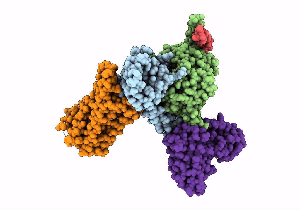
PDB ID:
7WU9
Keywords:
Title:
Cryo-EM structure of the human EP3-Gi signaling complex
Biological Source:
Source Organism(s):
Homo sapiens (Taxon ID: 9606)
Mus musculus (Taxon ID: 10090)
Mus musculus (Taxon ID: 10090)
Expression System(s):
Method Details:
Experimental Method:
Resolution:
3.38 Å
Aggregation State:
PARTICLE
Reconstruction Method:
SINGLE PARTICLE