7WU5 image
Entry Detail
PDB ID:
7WU5
EMDB ID:
Title:
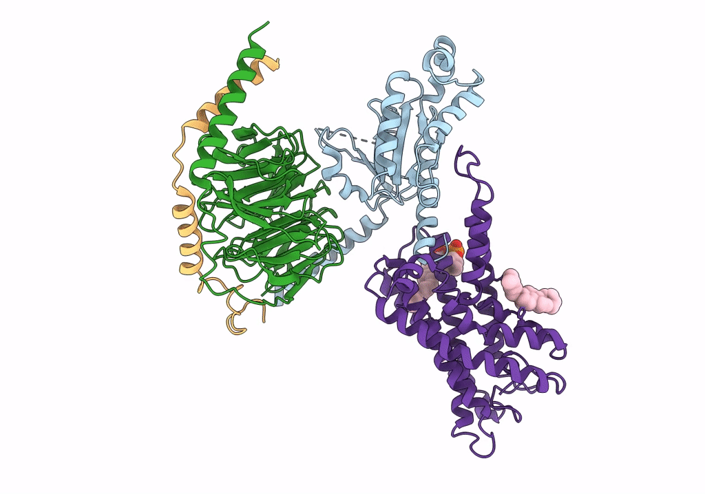
Cryo-EM structure of the adhesion GPCR ADGRF1(H565A/T567A) in complex with miniGi
Biological Source:
Source Organism:
Host Organism:
PDB Version:
Deposition Date:
2022-02-05
Release Date:
2022-04-27
Method Details:
Experimental Method:
Resolution:
3.00 Å
Aggregation State:
PARTICLE
Reconstruction Method:
SINGLE PARTICLE
Macromolecular Entities
Polymer Type:polypeptide(L)
Description:Guanine nucleotide-binding protein G(i) subunit alpha-1
Chain IDs:A
Chain Length:241
Number of Molecules:1
Biological Source:Homo sapiens
Polymer Type:polypeptide(L)
Description:Guanine nucleotide-binding protein G(I)/G(S)/G(T) subunit beta-1
Chain IDs:B
Chain Length:351
Number of Molecules:1
Biological Source:Homo sapiens
Polymer Type:polypeptide(L)
Description:Guanine nucleotide-binding protein G(I)/G(S)/G(O) subunit gamma-2
Chain IDs:C
Chain Length:71
Number of Molecules:1
Biological Source:Homo sapiens
Polymer Type:polypeptide(L)
Description:Adhesion G-protein coupled receptor F1
Mutations:H565A, T567A
Chain IDs:D (auth: R)
Chain Length:674
Number of Molecules:1
Biological Source:Homo sapiens
Primary Citation
Structural basis of tethered agonism of the adhesion GPCRs ADGRD1 and ADGRF1.
Nature 604 779 785 (2022)
PMID: 35418679 DOI: 10.1038/s41586-022-04580-w

Abstact

Adhesion G protein-coupled receptors (aGPCRs) are essential for a variety of physiological processes such as immune responses, organ development, cellular communication, proliferation and homeostasis1-7. An intrinsic manner of activation that involves a tethered agonist in the N-terminal region of the receptor has been proposed for the aGPCRs8,9, but its molecular mechanism remains elusive. Here we report the G protein-bound structures of ADGRD1 and ADGRF1, which exhibit many unique features with regard to the tethered agonism. The stalk region that proceeds the first transmembrane helix acts as the tethered agonist by forming extensive interactions with the transmembrane domain; these interactions are mostly conserved in ADGRD1 and ADGRF1, suggesting that a common stalk-transmembrane domain interaction pattern is shared by members of the aGPCR family. A similar stalk binding mode is observed in the structure of autoproteolysis-deficient ADGRF1, supporting a cleavage-independent manner of receptor activation. The stalk-induced activation is facilitated by a cascade of inter-helix interaction cores that are conserved in positions but show sequence variability in these two aGPCRs. Furthermore, the intracellular region of ADGRF1 contains a specific lipid-binding site, which proves to be functionally important and may serve as the recognition site for the previously discovered endogenous ADGRF1 ligand synaptamide. These findings highlight the diversity and complexity of the signal transduction mechanisms of the aGPCRs.

Legend

Protein

Chemical

Disease

Primary Citation of related structures