Deposition Date
2022-02-03
Release Date
2022-06-22
Last Version Date
2024-10-16
Entry Detail
PDB ID:
7WT4
Keywords:
Title:
Crystal structure of HLA-A*2402 complexed with 8-mer Influenza PB1 peptide
Biological Source:
Source Organism(s):
Homo sapiens (Taxon ID: 9606)
Influenza A virus (Taxon ID: 11320)
Influenza A virus (Taxon ID: 11320)
Expression System(s):
Method Details:
Experimental Method:
Resolution:
1.89 Å
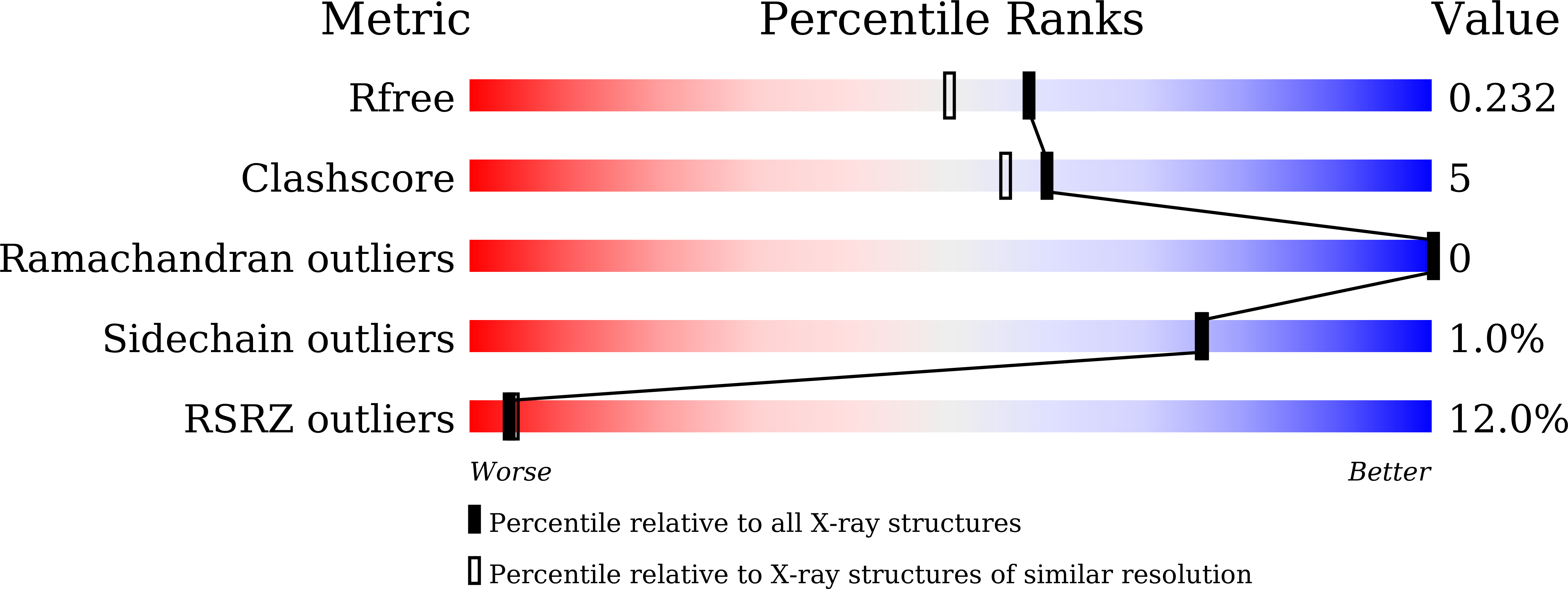
R-Value Free:
0.23
R-Value Work:
0.19
R-Value Observed:
0.20
Space Group:
P 1 21 1