7WPC image
Entry Detail
PDB ID:
7WPC
EMDB ID:
Keywords:
Title:
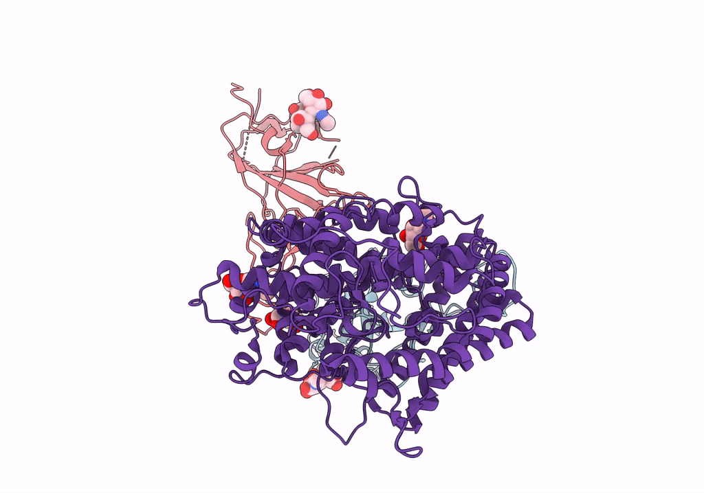
The second RBD of SARS-CoV-2 Omicron Variant in complexed with RBD-ACE2
Biological Source:
PDB Version:
Deposition Date:
2022-01-23
Release Date:
2022-03-09
Method Details:
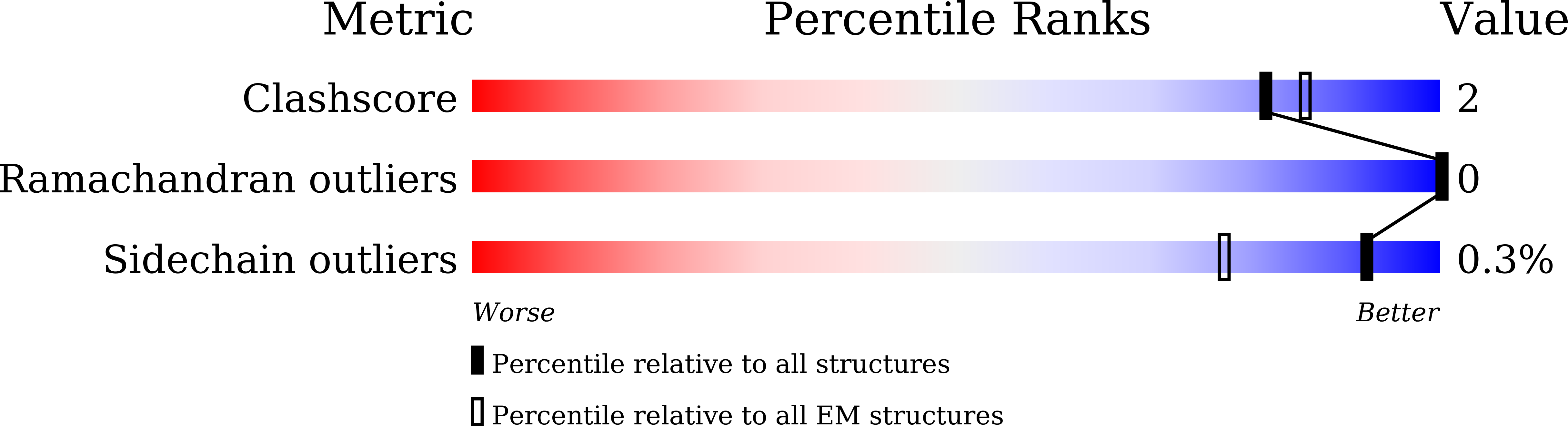
Experimental Method:
Resolution:
2.57 Å
Aggregation State:
PARTICLE
Reconstruction Method:
SINGLE PARTICLE
Macromolecular Entities
Polymer Type:polypeptide(L)
Description:Spike glycoprotein
Chain IDs:A, B
Chain Length:1205
Number of Molecules:2
Biological Source:Severe acute respiratory syndrome coronavirus 2
Polymer Type:polypeptide(L)
Description:Angiotensin-converting enzyme 2
Chain IDs:C (auth: D)
Chain Length:805
Number of Molecules:1
Biological Source:Homo sapiens
Primary Citation
Structures of the Omicron spike trimer with ACE2 and an anti-Omicron antibody.
Science 375 1048 1053 (2022)
PMID: 35133176 DOI: 10.1126/science.abn8863

Abstact

The severe acute respiratory syndrome coronavirus 2 (SARS-CoV-2) Omicron variant has become the dominant infective strain. We report the structures of the Omicron spike trimer on its own and in complex with angiotensin-converting enzyme 2 (ACE2) or an anti-Omicron antibody. Most Omicron mutations are located on the surface of the spike protein and change binding epitopes to many current antibodies. In the ACE2-binding site, compensating mutations strengthen receptor binding domain (RBD) binding to ACE2. Both the RBD and the apo form of the Omicron spike trimer are thermodynamically unstable. An unusual RBD-RBD interaction in the ACE2-spike complex supports the open conformation and further reinforces ACE2 binding to the spike trimer. A broad-spectrum therapeutic antibody, JMB2002, which has completed a phase 1 clinical trial, maintains neutralizing activity against Omicron. JMB2002 binds to RBD differently from other characterized antibodies and inhibits ACE2 binding.

Legend

Protein

Chemical

Disease

Primary Citation of related structures