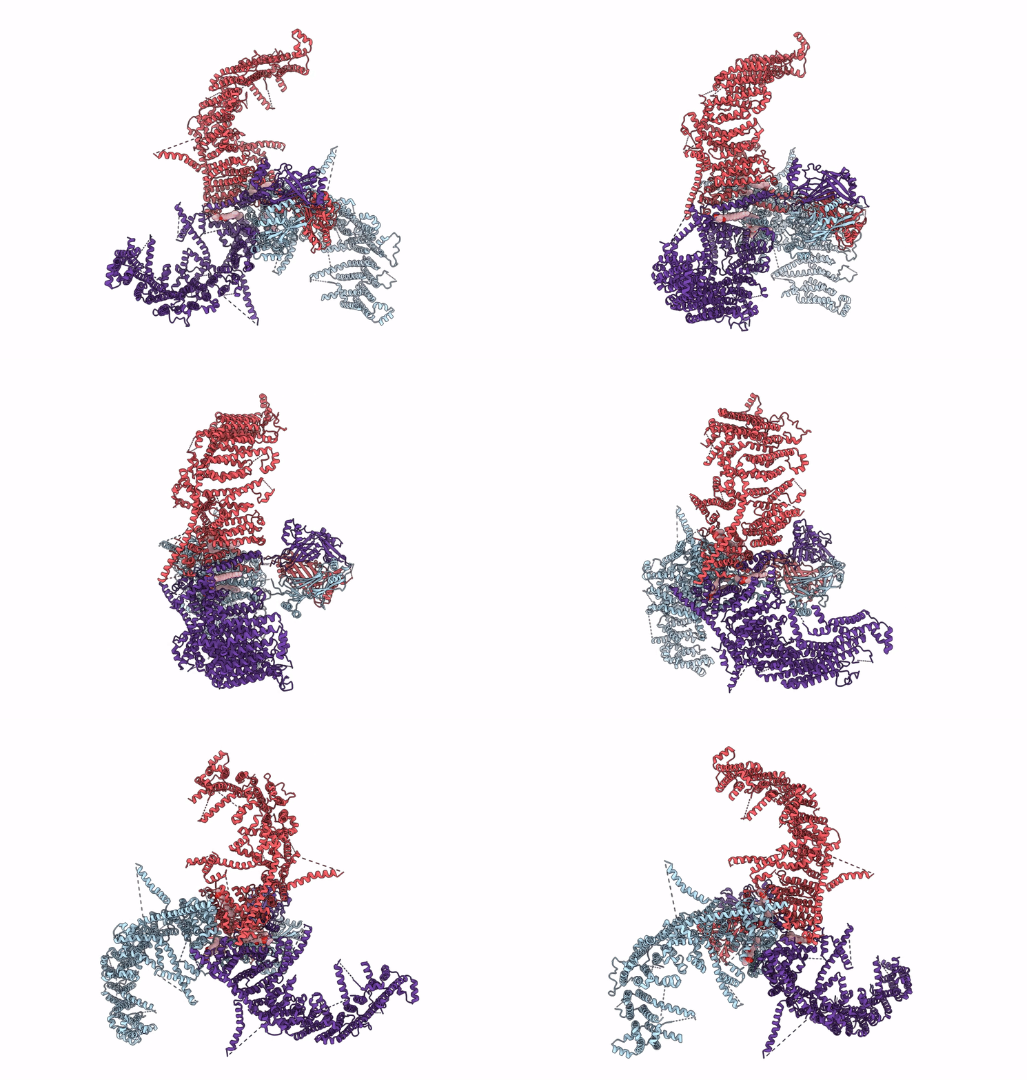
7WLU image
Deposition Date 2022-01-13
Release Date 2022-04-13
Last Version Date 2024-06-26
Entry Detail
PDB ID:
7WLU
Title:
The Flattened Structure of mPIEZO1 in Lipid Bilayer
Biological Source:
Source Organism:
Mus musculus (Taxon ID: 10090)
Host Organism:
Method Details:
Experimental Method:
Resolution:
6.81 Å
Aggregation State:
PARTICLE
Reconstruction Method:
SINGLE PARTICLE
Macromolecular Entities
Polymer Type:polypeptide(L)
Molecule:Piezo-type mechanosensitive ion channel component 1
Gene (Uniprot):Piezo1
Chain IDs:A, B (auth: C), C (auth: E)
Chain Length:2547
Number of Molecules:3
Biological Source:Mus musculus
Primary Citation
Structure deformation and curvature sensing of PIEZO1 in lipid membranes.
Nature 604 377 383 (2022)
PMID: 35388220 DOI: 10.1038/s41586-022-04574-8

Abstact

PIEZO channels respond to piconewton-scale forces to mediate critical physiological and pathophysiological processes1-5. Detergent-solubilized PIEZO channels form bowl-shaped trimers comprising a central ion-conducting pore with an extracellular cap and three curved and non-planar blades with intracellular beams6-10, which may undergo force-induced deformation within lipid membranes11. However, the structures and mechanisms underlying the gating dynamics of PIEZO channels in lipid membranes remain unresolved. Here we determine the curved and flattened structures of PIEZO1 reconstituted in liposome vesicles, directly visualizing the substantial deformability of the PIEZO1-lipid bilayer system and an in-plane areal expansion of approximately 300 nm2 in the flattened structure. The curved structure of PIEZO1 resembles the structure determined from detergent micelles, but has numerous bound phospholipids. By contrast, the flattened structure exhibits membrane tension-induced flattening of the blade, bending of the beam and detaching and rotating of the cap, which could collectively lead to gating of the ion-conducting pathway. On the basis of the measured in-plane membrane area expansion and stiffness constant of PIEZO1 (ref. 11), we calculate a half maximal activation tension of about 1.9 pN nm-1, matching experimentally measured values. Thus, our studies provide a fundamental understanding of how the notable deformability and structural rearrangement of PIEZO1 achieve exquisite mechanosensitivity and unique curvature-based gating in lipid membranes.

Legend

Protein

Chemical

Disease

Primary Citation of related structures