
Deposition Date
2021-12-14
Release Date
2022-01-12
Last Version Date
2024-11-13
Entry Detail
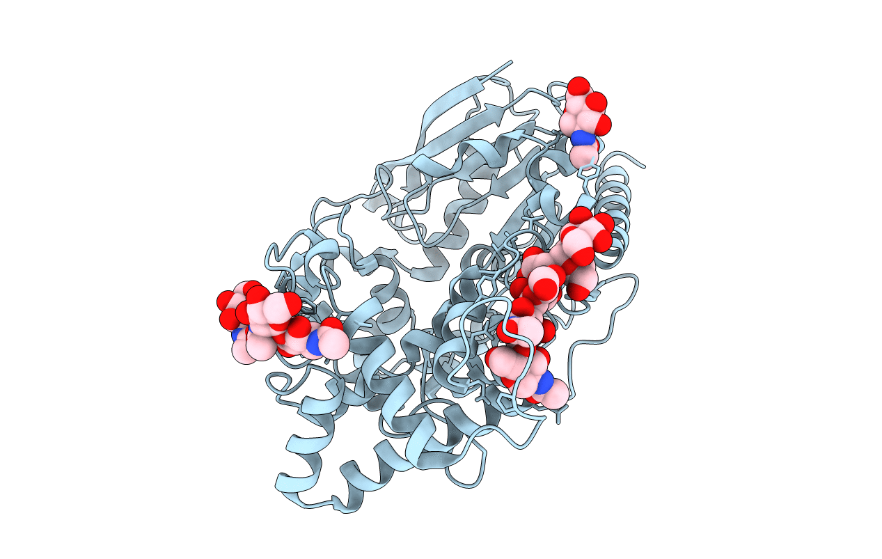
PDB ID:
7WAB
Keywords:
Title:
Crystal structure of the prolyl endoprotease, PEP, from Aspergillus niger
Biological Source:
Source Organism(s):
Aspergillus niger (Taxon ID: 5061)
Method Details:
Experimental Method:
Resolution:
1.75 Å
R-Value Free:
0.19
R-Value Work:
0.16
R-Value Observed:
0.16
Space Group:
P 4 21 2
