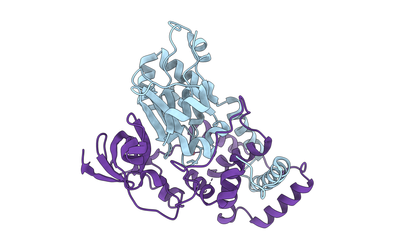
7W8L image
Entry Detail
PDB ID:
7W8L
Keywords:
Title:
Crystal Structure of Co-type nitrile hydratase mutant from Pseudonocardia thermophila - M46R
Biological Source:
PDB Version:
Deposition Date:
2021-12-08
Release Date:
2022-08-24
Method Details:
Experimental Method:
Resolution:
2.30 Å
R-Value Free:
0.23
R-Value Work:
0.17
R-Value Observed:
0.18
Space Group:
P 32 2 1
Macromolecular Entities
Polymer Type:polypeptide(L)
Description:Nitrile hydratase
Chain IDs:B (auth: A)
Chain Length:205
Number of Molecules:1
Biological Source:Pseudonocardia thermophila DSM 43832
Polymer Type:polypeptide(L)
Description:Cobalt-containing nitrile hydratase subunit beta
Mutations:M46R
Chain IDs:A (auth: B)
Chain Length:233
Number of Molecules:1
Biological Source:Pseudonocardia thermophila DSM 43832
Modified Residue
Compound ID Chain ID Parent Comp ID Details 2D Image
CSD B CYS modified residue
CSO B CYS modified residue
Ligand Molecules
Primary Citation
Insight into the broadened substrate scope of nitrile hydratase by static and dynamic structure analysis.
Chem Sci 13 8417 8428 (2022)
PMID: 35919716 DOI: 10.1039/d2sc02319a

Abstact

The narrow substrate scope limits the wide industrial application of enzymes. Here, we successfully broadened the substrate scope of a nitrile hydratase (NHase) through mutation of two tunnel entrance residues based on rational tunnel calculation. Two variants, with increased specific activity, especially toward bulky substrates, were obtained. Crystal structure analysis revealed that the mutations led to the expansion of the tunnel entrance, which might be conducive to substrate entry. More importantly, molecular dynamics simulations illustrated that the mutations introduced anti-correlated movements to the regions around the substrate tunnel and the active site, which would promote substrate access during the dynamic process of catalysis. Additionally, mutations on the corresponding tunnel entrance residues on other NHases also enhanced their activity toward bulky substrates. These results not only revealed that residues located at the enzyme surface were a key factor in enzyme catalytic performance, but also provided dynamic evidence for insight into enzyme substrate scope broadening.

Legend

Protein

Chemical

Disease

Primary Citation of related structures