7W70 image
Entry Detail
PDB ID:
7W70
Keywords:
Title:
Crystal structure of the PDZ-C domain fragment of Kangiella koreensis RseP orthologue
Biological Source:
Host Organism:
PDB Version:
Deposition Date:
2021-12-02
Release Date:
2022-09-07
Method Details:
Experimental Method:
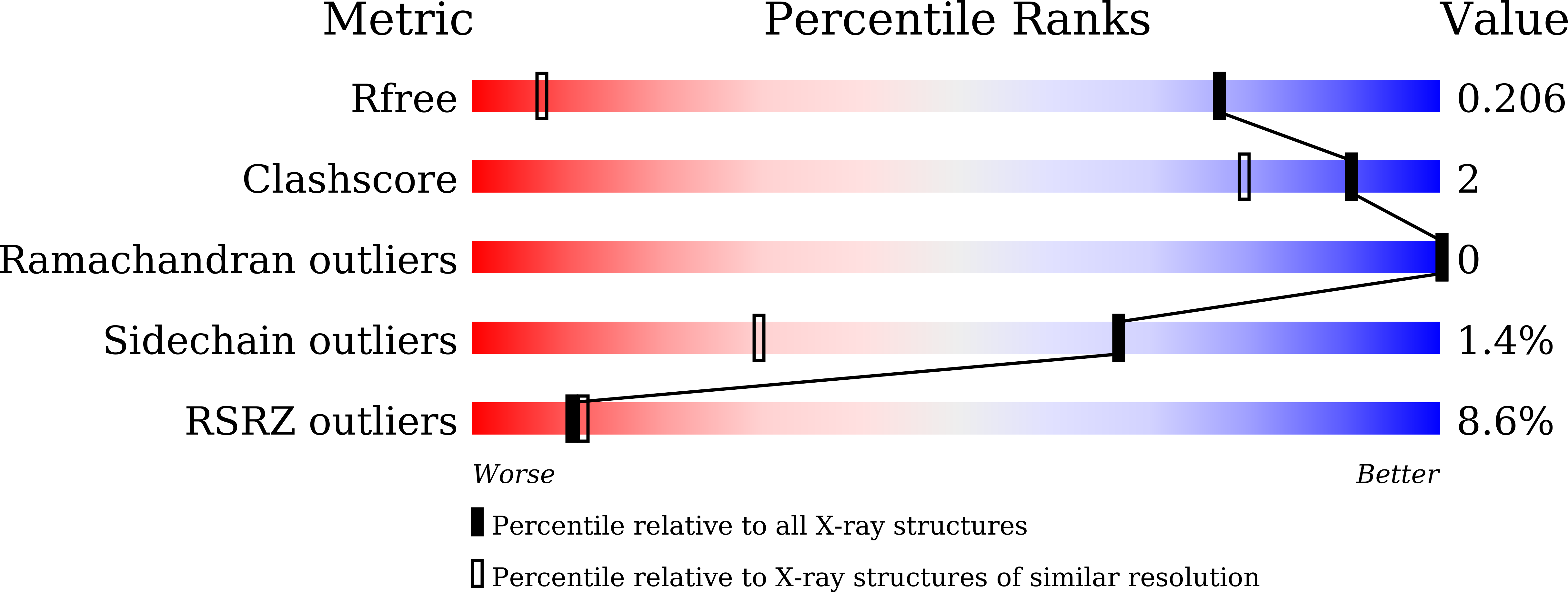
Resolution:
1.15 Å
R-Value Free:
0.20
R-Value Work:
0.19
R-Value Observed:
0.19
Space Group:
P 21 21 21
Macromolecular Entities
Polymer Type:polypeptide(L)
Description:Zinc metalloprotease
Chain IDs:A, B
Chain Length:87
Number of Molecules:2
Biological Source:Kangiella koreensis DSM 16069
Primary Citation
Mechanistic insights into intramembrane proteolysis by E. coli site-2 protease homolog RseP.
Sci Adv 8 eabp9011 eabp9011 (2022)
PMID: 36001659 DOI: 10.1126/sciadv.abp9011

Abstact

Site-2 proteases are a conserved family of intramembrane proteases that cleave transmembrane substrates to regulate signal transduction and maintain proteostasis. Here, we elucidated crystal structures of inhibitor-bound forms of bacterial site-2 proteases including Escherichia coli RseP. Structure-based chemical modification and cross-linking experiments indicated that the RseP domains surrounding the active center undergo conformational changes to expose the substrate-binding site, suggesting that RseP has a gating mechanism to regulate substrate entry. Furthermore, mutational analysis suggests that a conserved electrostatic linkage between the transmembrane and peripheral membrane-associated domains mediates the conformational changes. In vivo cleavage assays also support that the substrate transmembrane helix is unwound by strand addition to the intramembrane β sheet of RseP and is clamped by a conserved asparagine residue at the active center for efficient cleavage. This mechanism underlying the substrate binding, i.e., unwinding and clamping, appears common across distinct families of intramembrane proteases that cleave transmembrane segments.

Legend

Protein

Chemical

Disease

Primary Citation of related structures