
Deposition Date
2021-11-29
Release Date
2022-06-29
Last Version Date
2023-11-29
Entry Detail
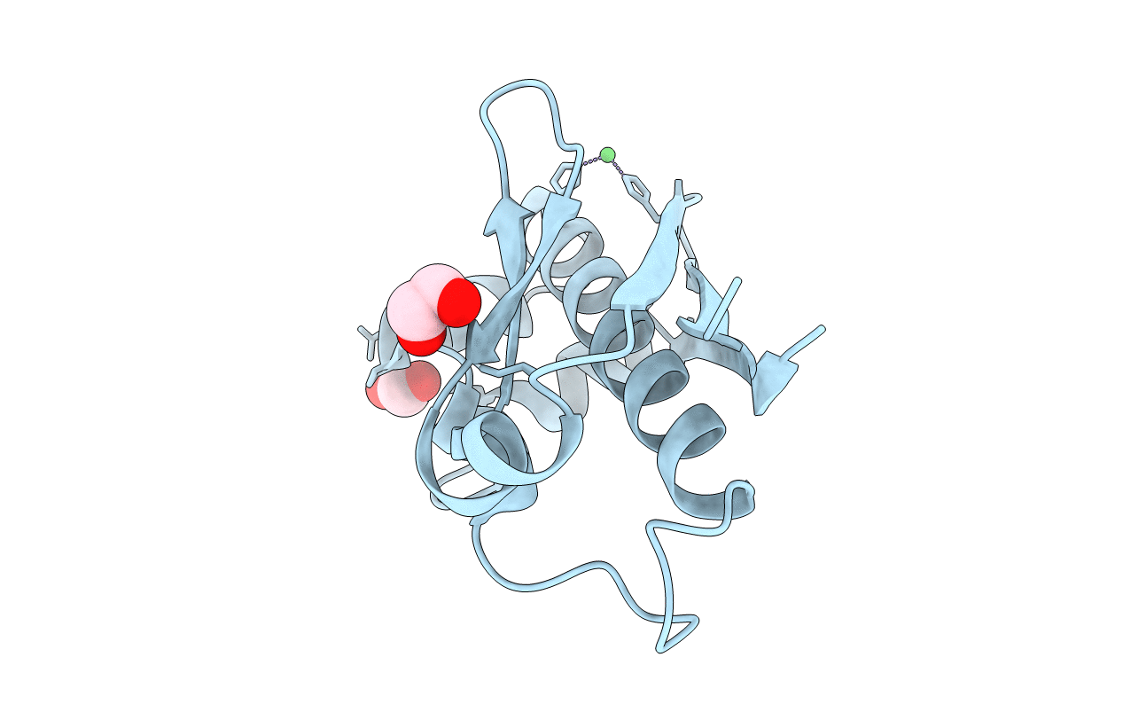
PDB ID:
7W58
Keywords:
Title:
Crystal structure of acyl-carrier protein synthase from Mycobacterium smegmatis
Biological Source:
Source Organism(s):
Mycolicibacterium smegmatis (Taxon ID: 1772)
Expression System(s):
Method Details:
Experimental Method:
Resolution:
2.27 Å
R-Value Free:
0.20
R-Value Work:
0.16
R-Value Observed:
0.16
Space Group:
H 3
