Deposition Date
2021-11-29
Release Date
2022-08-24
Last Version Date
2023-04-05
Entry Detail
Biological Source:
Source Organism(s):
Legionella pneumophila subsp. pneumophila str. Philadelphia 1 (Taxon ID: 272624)
Homo sapiens (Taxon ID: 9606)
Homo sapiens (Taxon ID: 9606)
Expression System(s):
Method Details:
Experimental Method:
Resolution:
2.64 Å
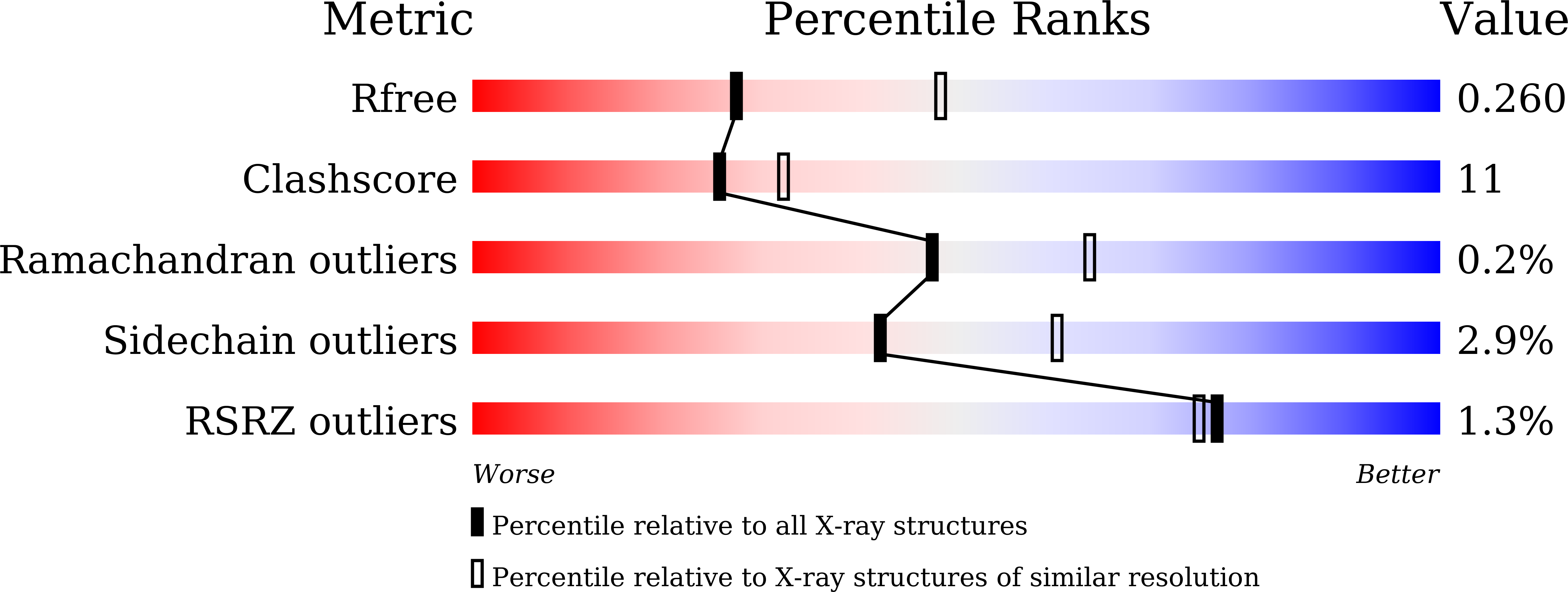
R-Value Free:
0.25
R-Value Work:
0.21
R-Value Observed:
0.21
Space Group:
P 21 21 21