7VZF image
Entry Detail
PDB ID:
7VZF
EMDB ID:
Keywords:
Title:
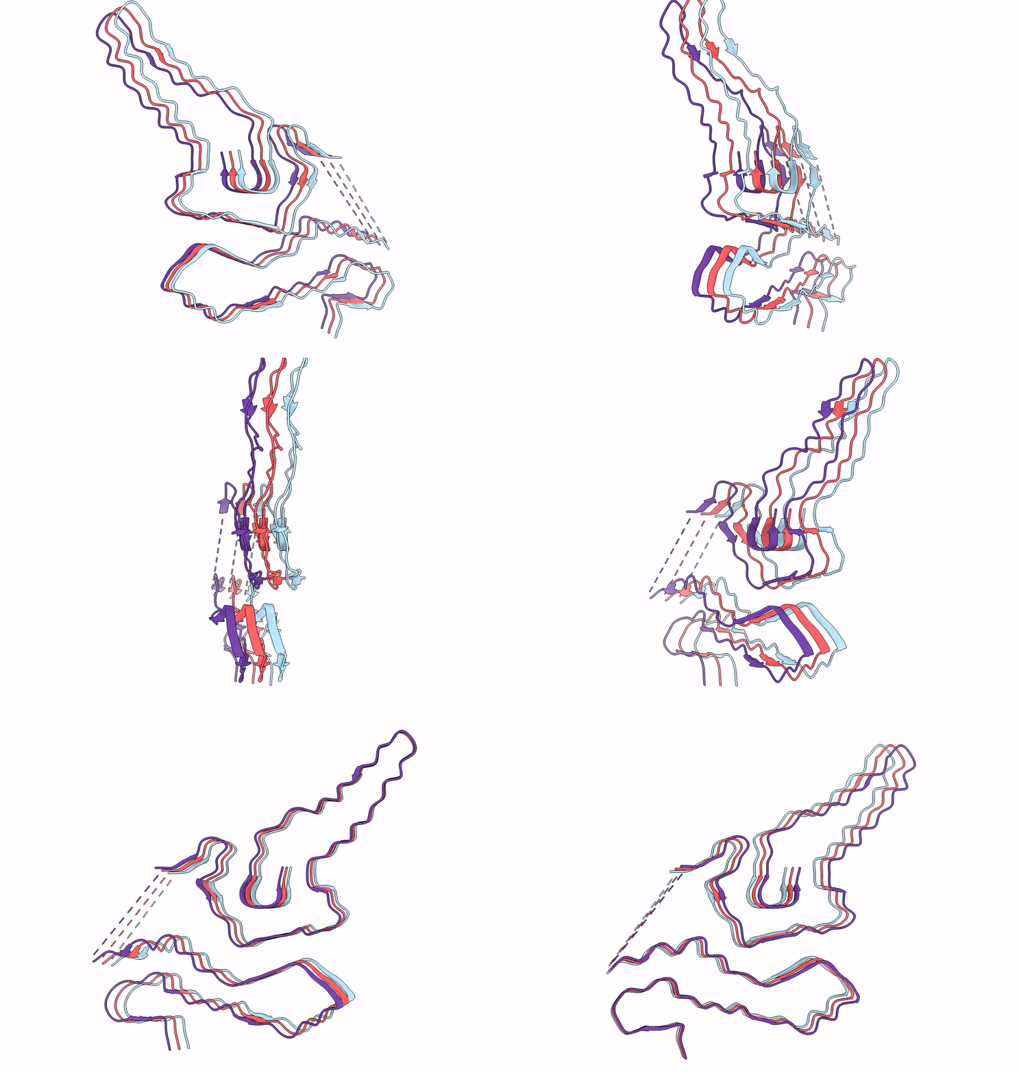
Cryo-EM structure of amyloid fibril formed by full-length human SOD1
Biological Source:
Source Organism:
Host Organism:
PDB Version:
Deposition Date:
2021-11-16
Release Date:
2022-06-29
Method Details:
Experimental Method:
Resolution:
2.95 Å
Aggregation State:
HELICAL ARRAY
Reconstruction Method:
HELICAL
Macromolecular Entities
Polymer Type:polypeptide(L)
Description:Superoxide dismutase [Cu-Zn]
Chain IDs:A, B, C
Chain Length:154
Number of Molecules:3
Biological Source:Homo sapiens
Ligand Molecules
Primary Citation
Cryo-EM structure of an amyloid fibril formed by full-length human SOD1 reveals its conformational conversion.
Nat Commun 13 3491 3491 (2022)
PMID: 35715417 DOI: 10.1038/s41467-022-31240-4

Abstact

Amyotrophic lateral sclerosis (ALS) is a neurodegenerative disease. Misfolded Cu, Zn-superoxide dismutase (SOD1) has been linked to both familial and sporadic ALS. SOD1 fibrils formed in vitro share toxic properties with ALS inclusions. Here we produced cytotoxic amyloid fibrils from full-length apo human SOD1 under reducing conditions and determined the atomic structure using cryo-EM. The SOD1 fibril consists of a single protofilament with a left-handed helix. The fibril core exhibits a serpentine fold comprising N-terminal segment (residues 3-55) and C-terminal segment (residues 86-153) with an intrinsic disordered segment. The two segments are zipped up by three salt bridge pairs. By comparison with the structure of apo SOD1 dimer, we propose that eight β-strands (to form a β-barrel) and one α-helix in the subunit of apo SOD1 convert into thirteen β-strands stabilized by five hydrophobic cavities in the SOD1 fibril. Our data provide insights into how SOD1 converts between structurally and functionally distinct states.

Legend

Protein

Chemical

Disease

Primary Citation of related structures