Deposition Date
2021-11-15
Release Date
2022-12-14
Last Version Date
2023-06-28
Entry Detail
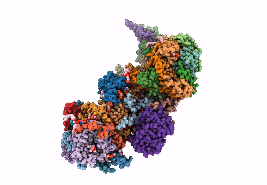
PDB ID:
7VYS
Keywords:
Title:
Membrane arm of active state CI from Q1-NADH dataset
Biological Source:
Source Organism(s):
Sus scrofa (Taxon ID: 9823)
Method Details:
Experimental Method:
Resolution:
2.50 Å
Aggregation State:
PARTICLE
Reconstruction Method:
SINGLE PARTICLE