Deposition Date
2021-11-09
Release Date
2022-08-10
Last Version Date
2024-11-13
Entry Detail
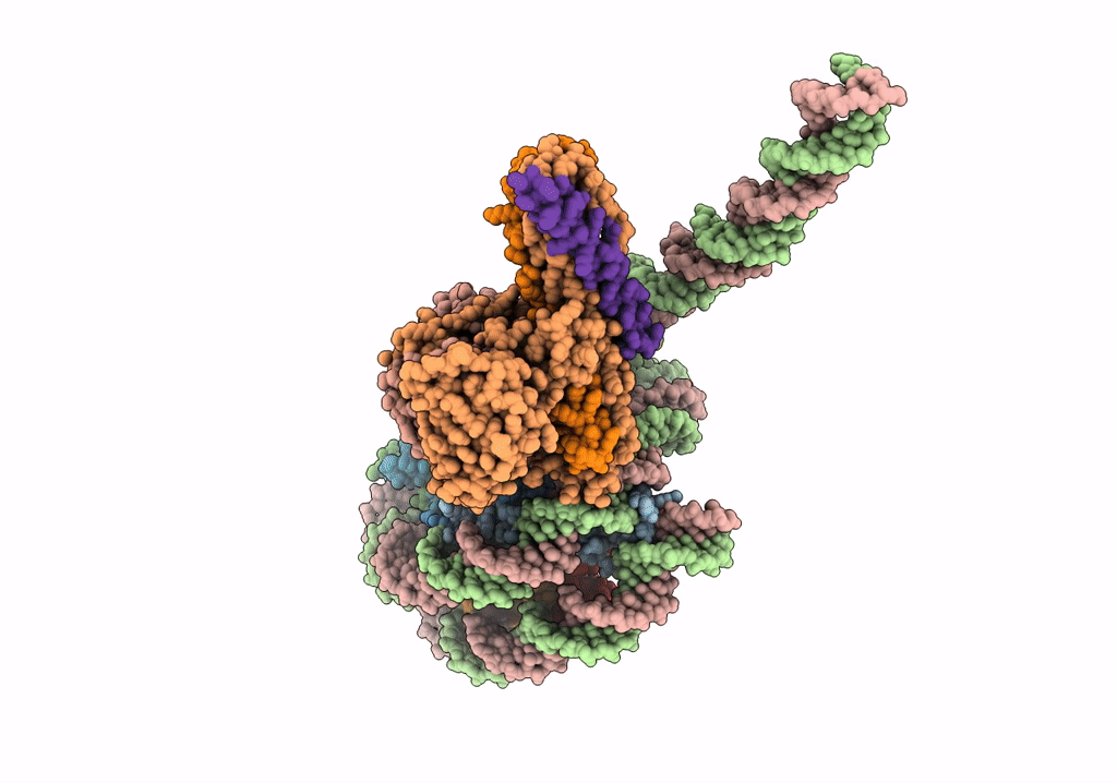
Biological Source:
Source Organism(s):
Xenopus laevis (Taxon ID: 8355)
Saccharomyces cerevisiae (Taxon ID: 4932)
Saccharomyces cerevisiae (Taxon ID: 4932)
Expression System(s):
Method Details:
Experimental Method:
Resolution:
3.40 Å
Aggregation State:
PARTICLE
Reconstruction Method:
SINGLE PARTICLE