7VVR image
Entry Detail
PDB ID:
7VVR
Keywords:
Title:
Bovine cytochrome c oxidese in CN-bound mixed valence state at 50 K
Biological Source:
Source Organism:
PDB Version:
Deposition Date:
2021-11-08
Release Date:
2022-11-16
Method Details:
Experimental Method:
Resolution:
1.65 Å
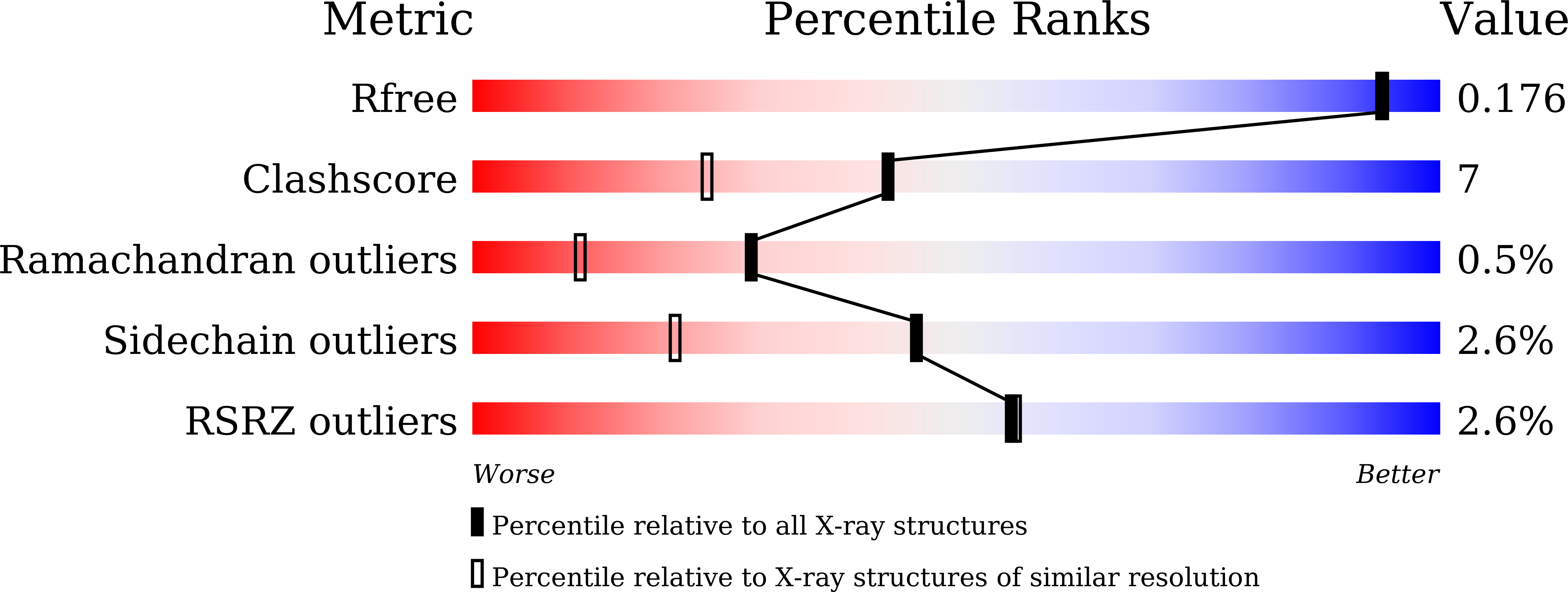
R-Value Free:
0.17
R-Value Work:
0.15
R-Value Observed:
0.15
Space Group:
P 21 21 21
Macromolecular Entities
Polymer Type:polypeptide(L)
Description:Cytochrome c oxidase subunit 1
Chain IDs:A, N
Chain Length:514
Number of Molecules:2
Biological Source:Bos taurus
Polymer Type:polypeptide(L)
Description:Cytochrome c oxidase subunit 2
Chain IDs:B, O
Chain Length:227
Number of Molecules:2
Biological Source:Bos taurus
Polymer Type:polypeptide(L)
Description:Cytochrome c oxidase subunit 3
Chain IDs:C, P
Chain Length:259
Number of Molecules:2
Biological Source:Bos taurus
Polymer Type:polypeptide(L)
Description:Cytochrome c oxidase subunit 4 isoform 1, mitochondrial
Chain IDs:D, Q
Chain Length:144
Number of Molecules:2
Biological Source:Bos taurus
Polymer Type:polypeptide(L)
Description:Cytochrome c oxidase subunit 5A, mitochondrial
Chain IDs:E, R
Chain Length:105
Number of Molecules:2
Biological Source:Bos taurus
Polymer Type:polypeptide(L)
Description:Cytochrome c oxidase subunit 5B, mitochondrial
Chain IDs:F, S
Chain Length:94
Number of Molecules:2
Biological Source:Bos taurus
Polymer Type:polypeptide(L)
Description:Cytochrome c oxidase subunit 6A2, mitochondrial
Chain IDs:G, T
Chain Length:84
Number of Molecules:2
Biological Source:Bos taurus
Polymer Type:polypeptide(L)
Description:Cytochrome c oxidase subunit 6B1
Chain IDs:H, U
Chain Length:79
Number of Molecules:2
Biological Source:Bos taurus
Polymer Type:polypeptide(L)
Description:Cytochrome c oxidase subunit 6C
Chain IDs:I, V
Chain Length:73
Number of Molecules:2
Biological Source:Bos taurus
Polymer Type:polypeptide(L)
Description:Cytochrome c oxidase subunit 7A1, mitochondrial
Chain IDs:J, W
Chain Length:58
Number of Molecules:2
Biological Source:Bos taurus
Polymer Type:polypeptide(L)
Description:Cytochrome c oxidase subunit 7B, mitochondrial
Chain IDs:K, X
Chain Length:49
Number of Molecules:2
Biological Source:Bos taurus
Polymer Type:polypeptide(L)
Description:Cytochrome c oxidase subunit 7C, mitochondrial
Chain IDs:L, Y
Chain Length:46
Number of Molecules:2
Biological Source:Bos taurus
Polymer Type:polypeptide(L)
Description:Cytochrome c oxidase subunit 8B, mitochondrial
Chain IDs:M, Z
Chain Length:43
Number of Molecules:2
Biological Source:Bos taurus
Modified Residue
Compound ID Chain ID Parent Comp ID Details 2D Image
FME A MET modified residue
SAC I SER modified residue
TPO G THR modified residue
Primary Citation
Crystallographic cyanide-probing for cytochrome c oxidase reveals structural bases suggesting that a putative proton transfer H-pathway pumps protons.
J.Biol.Chem. 299 105277 105277 (2023)
PMID: 37742916 DOI: 10.1016/j.jbc.2023.105277

Abstact

Cytochrome c oxidase (CcO) reduces O2 in the O2-reduction site by sequential four-electron donations through the low-potential metal sites (CuA and Fea). Redox-coupled X-ray crystal structural changes have been identified at five distinct sites including Asp51, Arg438, Glu198, the hydroxyfarnesyl ethyl group of heme a, and Ser382, respectively. These sites interact with the putative proton-pumping H-pathway. However, the metal sites responsible for each structural change have not been identified, since these changes were detected as structural differences between the fully reduced and fully oxidized CcOs. Thus, the roles of these structural changes in the CcO function are yet to be revealed. X-ray crystal structures of cyanide-bound CcOs under various oxidation states showed that the O2-reduction site controlled only the Ser382-including site, while the low-potential metal sites induced the other changes. This finding indicates that these low-potential site-inducible structural changes are triggered by sequential electron-extraction from the low-potential sites by the O2-reduction site and that each structural change is insensitive to the oxidation and ligand-binding states of the O2-reduction site. Because the proton/electron coupling efficiency is constant (1:1), regardless of the reaction progress in the O2-reduction site, the structural changes induced by the low-potential sites are assignable to those critically involved in the proton pumping, suggesting that the H-pathway, facilitating these low-potential site-inducible structural changes, pumps protons. Furthermore, a cyanide-bound CcO structure suggests that a hypoxia-inducible activator, Higd1a, activates the O2-reduction site without influencing the electron transfer mechanism through the low-potential sites, kinetically confirming that the low-potential sites facilitate proton pump.

Legend

Protein

Chemical

Disease

Primary Citation of related structures