7VRX image
Entry Detail
PDB ID:
7VRX
Keywords:
Title:
Pad-1 in the absence of substrate
Biological Source:
Source Organism:
Host Organism:
PDB Version:
Deposition Date:
2021-10-25
Release Date:
2022-04-20
Method Details:
Experimental Method:
Resolution:
1.97 Å
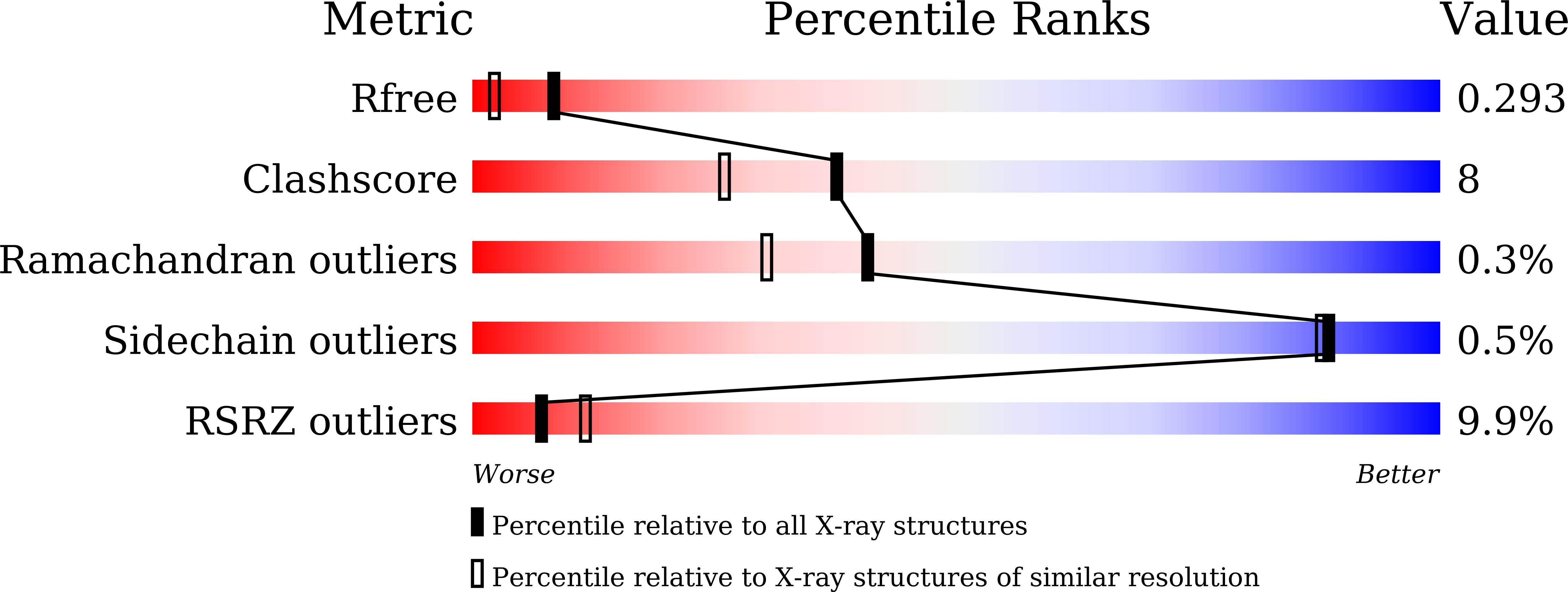
R-Value Free:
0.29
R-Value Work:
0.25
R-Value Observed:
0.25
Space Group:
P 65
Macromolecular Entities
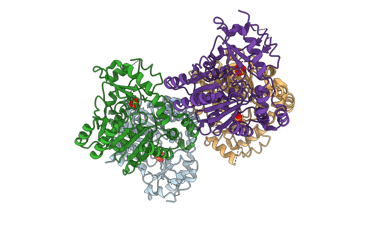
Polymer Type:polypeptide(L)
Description:Aminotransferase
Chain IDs:A, B, C, D
Chain Length:404
Number of Molecules:4
Biological Source:Solanum melongena
Modified Residue
Compound ID Chain ID Parent Comp ID Details 2D Image
LLP A LYS modified residue
Ligand Molecules
Primary Citation
Structural and biochemical basis for the substrate specificity of Pad-1, an indole-3-pyruvic acid aminotransferase in auxin homeostasis.
J.Struct.Biol. 214 107857 107857 (2022)
PMID: 35395410 DOI: 10.1016/j.jsb.2022.107857

Abstact

Phytohormone indole-3-acetic acid (IAA) plays a vital role in regulating plant growth and development. Tryptophan-dependent IAA biosynthesis participates in IAA homeostasis by producing IAA via two sequential reactions, which involve a conversion of tryptophan to indole-3-pyruvic acid (IPyA) by tryptophan aminotransferase (TAA1) followed by the irreversible formation of IAA in the second reaction. Pad-1 from Solanaceae plants regulates IAA levels by catalyzing a reverse reaction of the first step of IAA biosynthesis. Pad-1 is a pyridoxal phosphate (PLP)-dependent aminotransferase, with IPyA as the amino acceptor and l-glutamine as the amino donor. Currently, the structural and functional basis for the substrate specificity of Pad-1 remains poorly understood. In this study, we carried out structural and kinetic analyses of Pad-1 from Solanum melongena. Pad-1 is a homodimeric enzyme, with coenzyme PLP present between a central large α/β domain and a protruding small domain. The active site of Pad-1 includes a vacancy near the phosphate group (P-side) and the 3'-O (O-side) of PLP. These features are distinct from those of TAA1, which is homologous in an overall structure with Pad-1 but includes only the P-side region in the active site. Kinetic analysis suggests that P-side residues constitute a binding pocket for l-glutamine, and O-side residues of Phe124 and Ile350 are involved in the binding of IPyA. These studies illuminate distinct differences in the active site between Pad-1 and TAA1, and provide structural and functional insights into the substrate specificity of Pad-1.

Legend

Protein

Chemical

Disease

Primary Citation of related structures