7VR5 image
Entry Detail
PDB ID:
7VR5
Title:
Crystal structure of CmABCB1 W114Y/W161Y/W363Y/W364Y/M391W (4WY/M391W) mutant
Biological Source:
PDB Version:
Deposition Date:
2021-10-21
Release Date:
2022-09-07
Method Details:
Experimental Method:
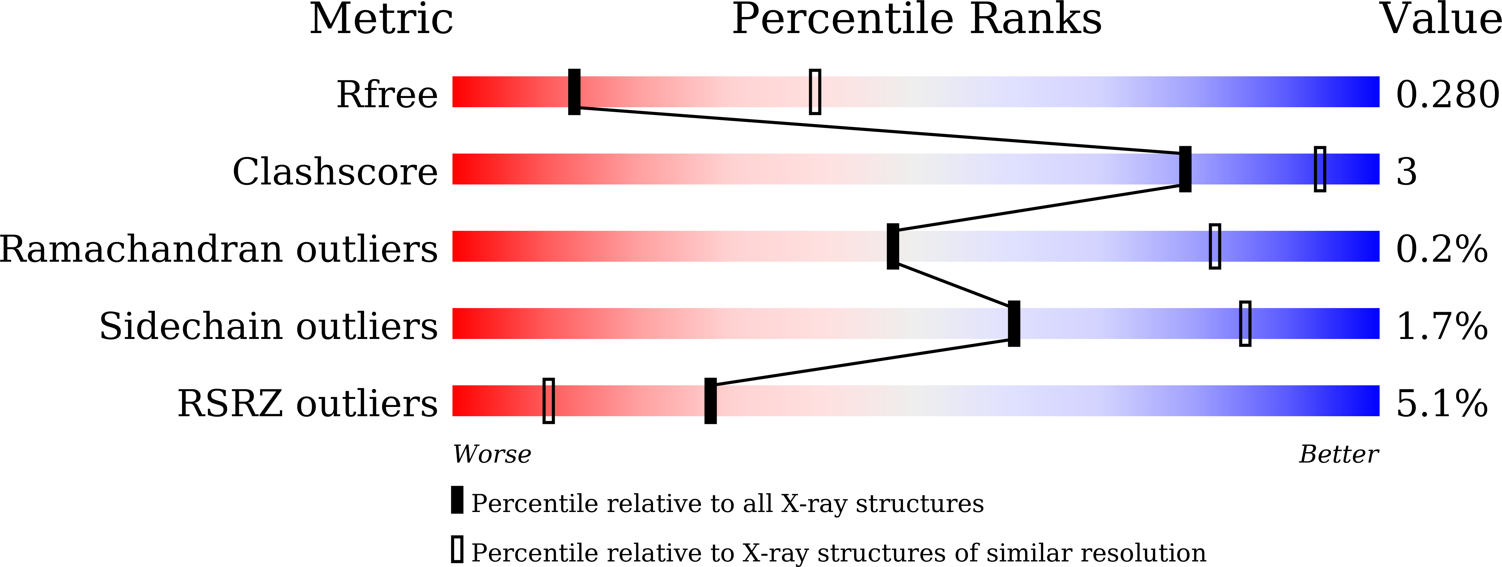
Resolution:
3.00 Å
R-Value Free:
0.26
R-Value Work:
0.21
R-Value Observed:
0.21
Space Group:
H 3 2
Macromolecular Entities
Polymer Type:polypeptide(L)
Description:Probable ATP-dependent transporter ycf16
Mutations:W114Y,W161Y,W363Y,W364Y,M391W
Chain IDs:A
Chain Length:612
Number of Molecules:1
Biological Source:Cyanidioschyzon merolae (strain 10D)
Primary Citation
Structure-based alteration of tryptophan residues of the multidrug transporter CmABCB1 to assess substrate binding using fluorescence spectroscopy.
Protein Sci. 31 e4331 e4331 (2022)
PMID: 35634783 DOI: 10.1002/pro.4331

Abstact

ABCB1, also known as P-glycoprotein, is an essential component of many physiological barriers and extrudes a variety of hydrophobic chemicals out of the cell. Structures of ABCB1 provided insights into the structural changes that occur upon ATP binding and the characteristic architecture of the substrate binding site. Yet, the structure-function relationship between substrate binding and transporting still remains largely obscured because there is no robust method for accurately measuring substrate binding constants. The methods currently used cannot identify whether the bound substrates are located in the inner chamber of the molecule in the transmembrane region or not because of the low spatial resolution. Here, we report a system for measuring the affinity of substrate binding to the Cyanidioschyzon merolae ABCB1 (CmABCB1) using site-specific tryptophan (Trp) fluorescence quenching. We designed a CmABCB1 mutant with an extrinsic Trp residue introduced into the inner chamber. Trp fluorescence was quenched by three substrates and one inhibitor, including rhodamine 6G, in a saturable fashion, allowing for accurate estimation of the dissociation constant (KD) for each molecule. The KD for rhodamine 6G is similar to that determined using a reciprocal fluorescence quenching assay using rhodamine 6G fluorescence, suggesting that Trp fluorescence of the mutant was quenched by the interaction between the extrinsic Trp and substrates bound in the inner chamber. Structural comparison of the ABCB1 structures suggests that the system presented in this study could be ideal method of choice to determine the substrate binding affinities of compounds bound to the chamber of mammalian ABCB1.

Legend

Protein

Chemical

Disease

Primary Citation of related structures