7VOK image
Deposition Date 2021-10-14
Release Date 2022-10-19
Last Version Date 2024-04-24
Entry Detail
PDB ID:
7VOK
Title:
The Crystal structure of EF-Tu and GDP from Mycobacterium tuberculosis
Biological Source:
Source Organism:
Host Organism:
Method Details:
Experimental Method:
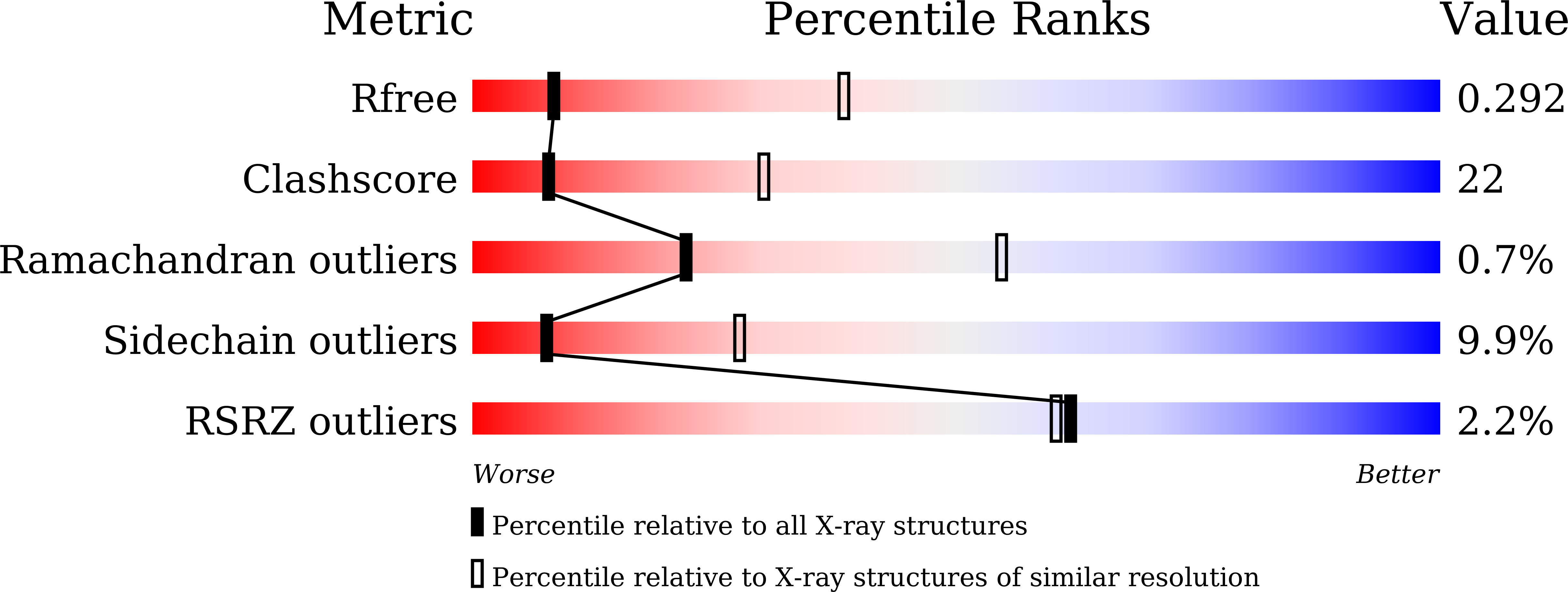
Resolution:
3.40 Å
R-Value Free:
0.30
R-Value Work:
0.24
R-Value Observed:
0.24
Space Group:
P 1 21 1
Macromolecular Entities
Structures with similar UniProt ID
Protein Blast
Polymer Type:polypeptide(L)
Molecule:Elongation factor Tu
Chain IDs:A, B, C, D
Chain Length:396
Number of Molecules:4
Biological Source:Mycobacterium tuberculosis
Primary Citation
Structural insights of the elongation factor EF-Tu complexes in protein translation of Mycobacterium tuberculosis.
Commun Biol 5 1052 1052 (2022)
PMID: 36192483 DOI: 10.1038/s42003-022-04019-y

Abstact

Tuberculosis (TB) caused by Mycobacterium tuberculosis (Mtb) is the second-deadliest infectious disease worldwide. Emerging evidence shows that the elongation factor EF-Tu could be an excellent target for treating Mtb infection. Here, we report the crystal structures of Mtb EF-Tu•EF-Ts and EF-Tu•GDP complexes, showing the molecular basis of EF-Tu's representative recycling and inactive forms in protein translation. Mtb EF-Tu binds with EF-Ts at a 1:1 ratio in solution and crystal packing. Mutation and SAXS analysis show that EF-Ts residues Arg13, Asn82, and His149 are indispensable for the EF-Tu/EF-Ts complex formation. The GDP binding pocket of EF-Tu dramatically changes conformations upon binding with EF-Ts, sharing a similar GDP-exchange mechanism in E. coli and T. ther. Also, the FDA-approved drug Osimertinib inhibits the growth of M. smegmatis, H37Ra, and M. bovis BCG strains by directly binding with EF-Tu. Thus, our work reveals the structural basis of Mtb EF-Tu in polypeptide synthesis and may provide a promising candidate for TB treatment.

Legend

Protein

Chemical

Disease

Primary Citation of related structures
Feedback Form
Name
Email
Institute
Feedback