7VNS image
Deposition Date 2021-10-12
Release Date 2022-05-25
Last Version Date 2024-11-13
Entry Detail
PDB ID:
7VNS
Title:
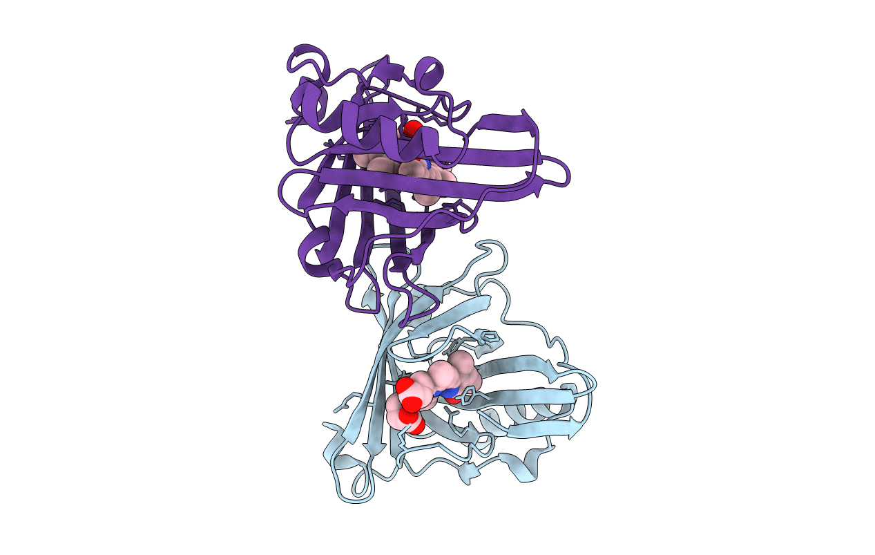
Sandercyanin mutant E79A-Biliverdin complex
Biological Source:
Source Organism:
Host Organism:
Method Details:
Experimental Method:
Resolution:
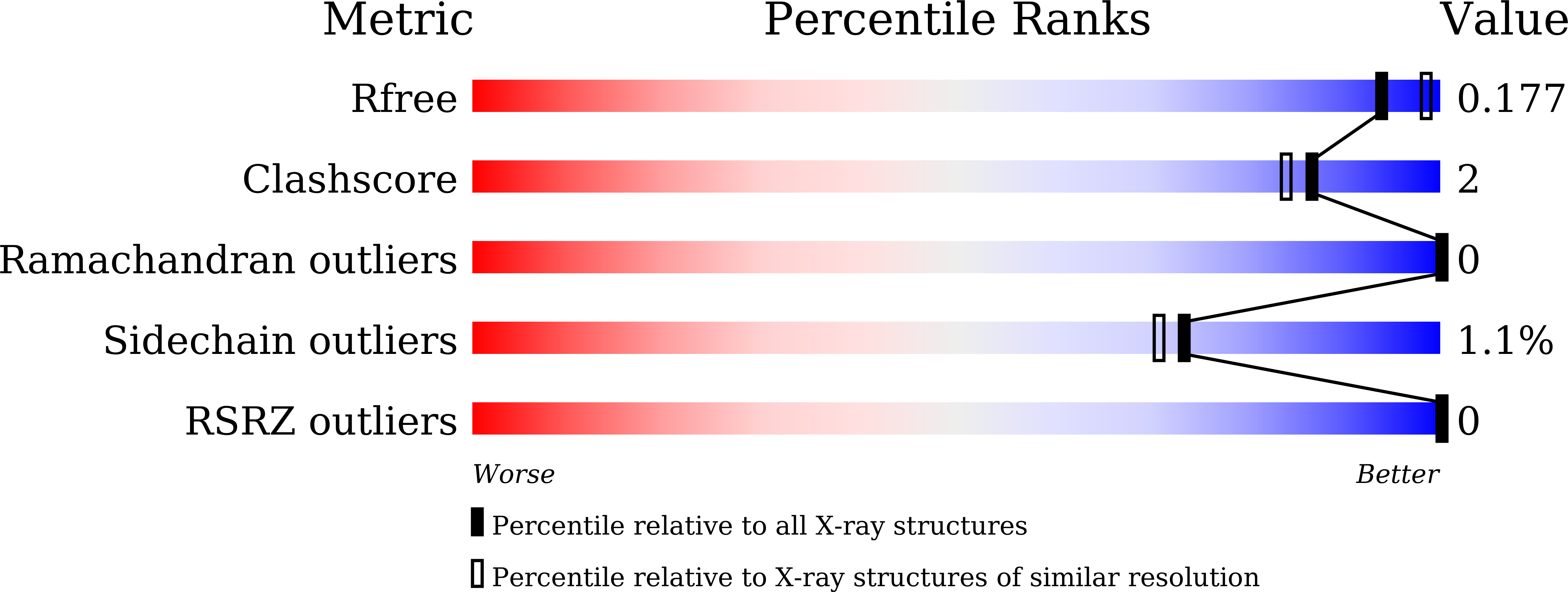
1.95 Å
R-Value Free:
0.17
R-Value Work:
0.17
R-Value Observed:
0.17
Space Group:
P 63 2 2
Macromolecular Entities
Structures with similar UniProt ID
Protein Blast
Polymer Type:polypeptide(L)
Molecule:Sandercyanin Fluorescent Protein
Mutagens:E79A
Chain IDs:A, B
Chain Length:183
Number of Molecules:2
Biological Source:Sander vitreus
Ligand Molecules
Primary Citation
Phenylalanine stacking enhances the red fluorescence of biliverdin IX alpha on UV excitation in sandercyanin fluorescent protein.
Febs Lett. 596 796 805 (2022)
PMID: 35020202 DOI: 10.1002/1873-3468.14281

Abstact

Biliverdin IXα (BV) binds to several prokaryotic and eukaryotic proteins. How nature exploits the versatility of BV's properties is not fully understood. Unlike free BV, the Sandercyanin fluorescent protein bound to BV (SFP-BV) shows enhanced red fluorescence (675 nm) on excitation in the UV region (380 nm). Site-directed mutagenesis showed that the BV complex of two SFP variants, F55A and E79A, resulted in the loss of red fluorescence. Crystal structures of the complexes of these proteins with BV show the absence of stacking interactions of the F55 phenyl ring with BV. BV changes from ZZZssa conformation in the wild-type to ZZZsss conformation in the variants. In the nonfluorescent mutants, the lowest excited state is destabilized, resulting in nonradiative decay.

Legend

Protein

Chemical

Disease

Primary Citation of related structures