7VJ7 image
Deposition Date 2021-09-28
Release Date 2022-03-09
Last Version Date 2023-11-29
Entry Detail
PDB ID:
7VJ7
Keywords:
Title:
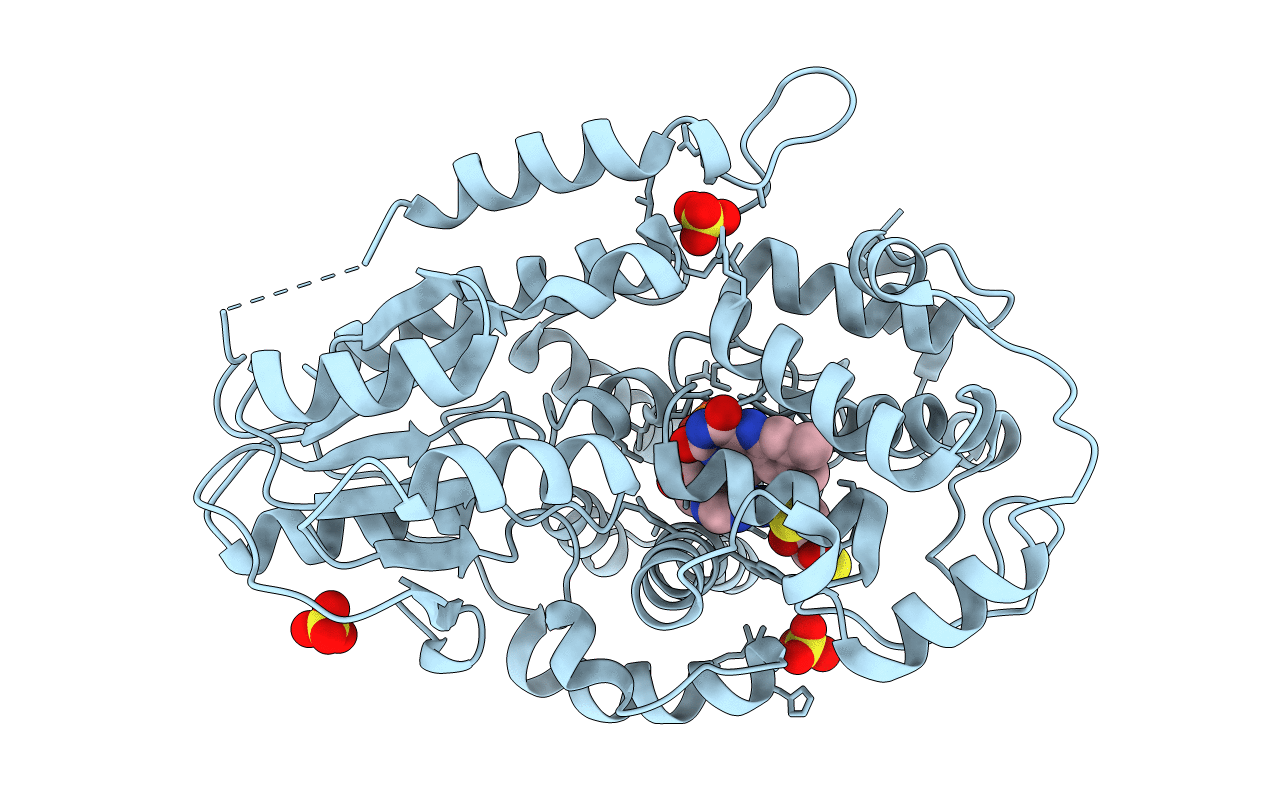
SFX structure of archaeal class II CPD photolyase from Methanosarcina mazei in the fully reduced state
Biological Source:
Source Organism:
Method Details:
Experimental Method:
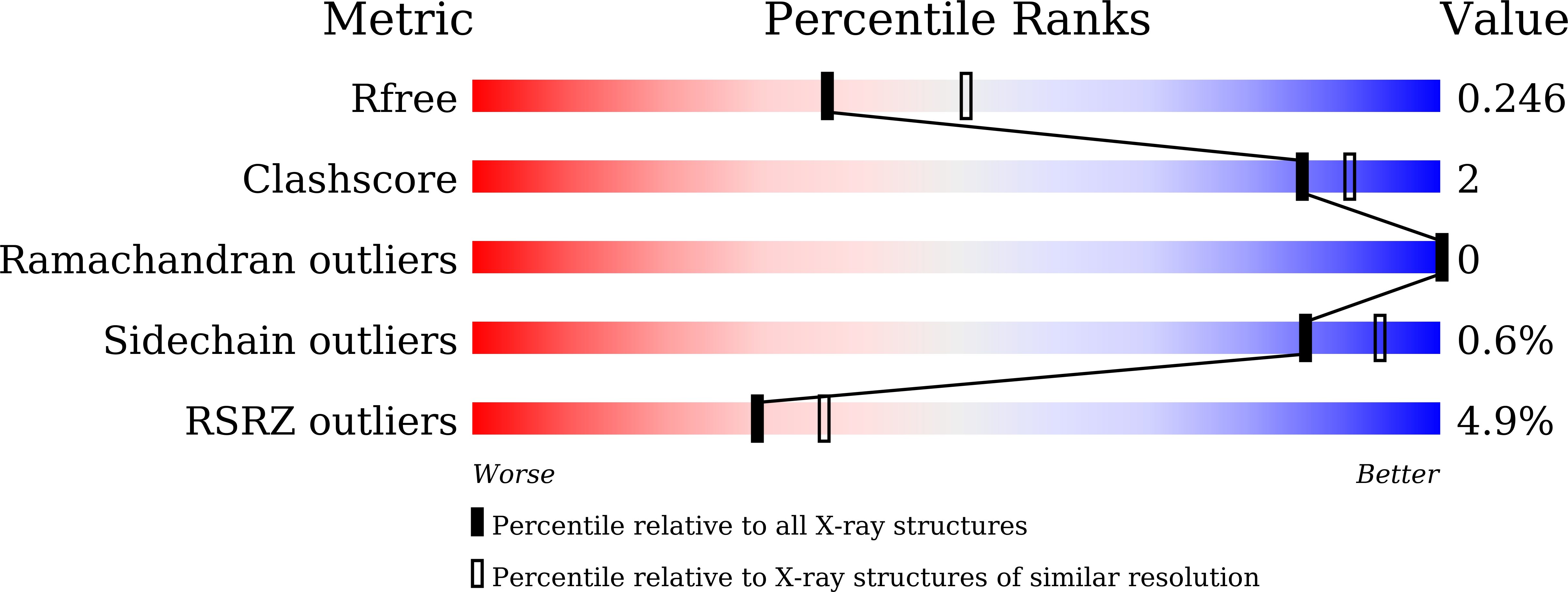
Resolution:
2.30 Å
R-Value Free:
0.25
R-Value Work:
0.21
R-Value Observed:
0.21
Space Group:
P 43 21 2
Macromolecular Entities
Structures with similar UniProt ID
Protein Blast
Polymer Type:polypeptide(L)
Molecule:DNA photolyase
Gene (Uniprot):MM_0852
Mutagens:M377T
Chain IDs:A
Chain Length:482
Number of Molecules:1
Biological Source:Methanosarcina mazei Go1
Primary Citation

Abstact

Flavin coenzymes are universally found in biological redox reactions. DNA photolyases, with their flavin chromophore (FAD), utilize blue light for DNA repair and photoreduction. The latter process involves two single-electron transfers to FAD with an intermittent protonation step to prime the enzyme active for DNA repair. Here we use time-resolved serial femtosecond X-ray crystallography to describe how light-driven electron transfers trigger subsequent nanosecond-to-microsecond entanglement between FAD and its Asn/Arg-Asp redox sensor triad. We found that this key feature within the photolyase-cryptochrome family regulates FAD re-hybridization and protonation. After first electron transfer, the FAD•- isoalloxazine ring twists strongly when the arginine closes in to stabilize the negative charge. Subsequent breakage of the arginine-aspartate salt bridge allows proton transfer from arginine to FAD•-. Our molecular videos demonstrate how the protein environment of redox cofactors organizes multiple electron/proton transfer events in an ordered fashion, which could be applicable to other redox systems such as photosynthesis.

Legend

Protein

Chemical

Disease

Primary Citation of related structures