Deposition Date
2021-09-26
Release Date
2022-09-28
Last Version Date
2024-10-23
Entry Detail
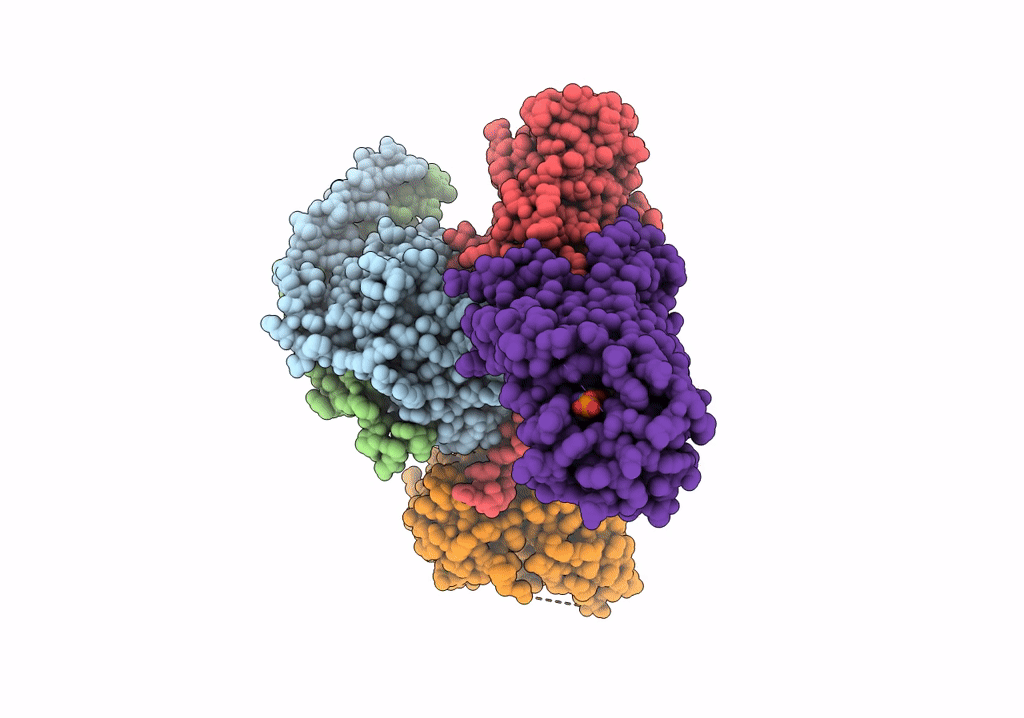
PDB ID:
7VIE
Keywords:
Title:
Cryo-EM structure of Gi coupled Sphingosine 1-phosphate receptor bound with S1P
Biological Source:
Source Organism(s):
Homo sapiens (Taxon ID: 9606)
Mus musculus (Taxon ID: 10090)
Mus musculus (Taxon ID: 10090)
Expression System(s):
Method Details:
Experimental Method:
Resolution:
2.86 Å
Aggregation State:
PARTICLE
Reconstruction Method:
SINGLE PARTICLE