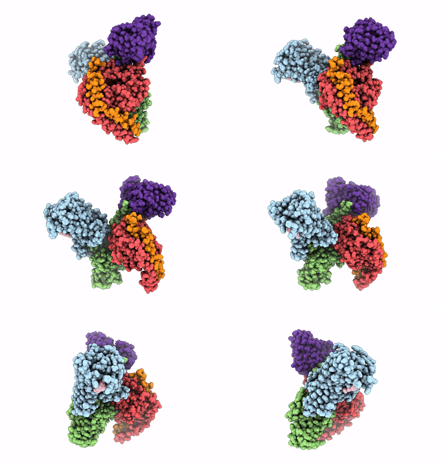
7VGZ image
Entry Detail
PDB ID:
7VGZ
EMDB ID:
Title:
MT1-remalteon-Gi complex
Biological Source:
PDB Version:
Deposition Date:
2021-09-20
Release Date:
2022-03-02
Method Details:
Experimental Method:
Resolution:
3.30 Å
Aggregation State:
PARTICLE
Reconstruction Method:
SINGLE PARTICLE
Macromolecular Entities
Polymer Type:polypeptide(L)
Description:Melatonin receptor type 1A
Chain IDs:A (auth: B)
Chain Length:350
Number of Molecules:1
Biological Source:Homo sapiens
Polymer Type:polypeptide(L)
Description:Guanine nucleotide-binding protein G(i) subunit alpha-1
Chain IDs:B (auth: C)
Chain Length:353
Number of Molecules:1
Biological Source:Homo sapiens
Polymer Type:polypeptide(L)
Description:Guanine nucleotide-binding protein G(I)/G(S)/G(T) subunit beta-1
Chain IDs:C (auth: D)
Chain Length:345
Number of Molecules:1
Biological Source:Rattus norvegicus
Polymer Type:polypeptide(L)
Description:Guanine nucleotide-binding protein G(I)/G(S)/G(O) subunit gamma-2
Chain IDs:D (auth: E)
Chain Length:67
Number of Molecules:1
Biological Source:Bos taurus
Polymer Type:polypeptide(L)
Description:scFv16
Chain IDs:E (auth: F)
Chain Length:256
Number of Molecules:1
Biological Source:synthetic construct
Primary Citation
Structural basis of the ligand binding and signaling mechanism of melatonin receptors.
Nat Commun 13 454 454 (2022)
PMID: 35075127 DOI: 10.1038/s41467-022-28111-3

Abstact

Melatonin receptors (MT1 and MT2 in humans) are family A G protein-coupled receptors that respond to the neurohormone melatonin to regulate circadian rhythm and sleep. Numerous efforts have been made to develop drugs targeting melatonin receptors for the treatment of insomnia, circadian rhythm disorder, and cancer. However, designing subtype-selective melatonergic drugs remains challenging. Here, we report the cryo-EM structures of the MT1-Gi signaling complex with 2-iodomelatonin and ramelteon and the MT2-Gi signaling complex with ramelteon. These structures, together with the reported functional data, reveal that although MT1 and MT2 possess highly similar orthosteric ligand-binding pockets, they also display distinctive features that could be targeted to design subtype-selective drugs. The unique structural motifs in MT1 and MT2 mediate structural rearrangements with a particularly wide opening on the cytoplasmic side. Gi is engaged in the receptor core shared by MT1 and MT2 and presents a conformation deviating from those in other Gi complexes. Together, our results provide new clues for designing melatonergic drugs and further insights into understanding the G protein coupling mechanism.

Legend

Protein

Chemical

Disease

Primary Citation of related structures