
Deposition Date
2021-09-13
Release Date
2021-11-03
Last Version Date
2025-07-02
Entry Detail
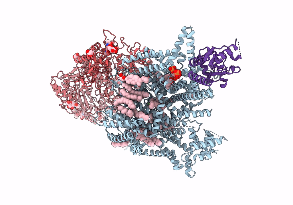
PDB ID:
7VFS
Keywords:
Title:
Human N-type voltage gated calcium channel CaV2.2-alpha2/delta1-beta1 complex, apo state
Biological Source:
Source Organism(s):
Homo sapiens (Taxon ID: 9606)
Expression System(s):
Method Details:
Experimental Method:
Resolution:
2.80 Å
Aggregation State:
PARTICLE
Reconstruction Method:
SINGLE PARTICLE
