
Deposition Date
2021-09-07
Release Date
2021-09-22
Last Version Date
2025-09-17
Entry Detail
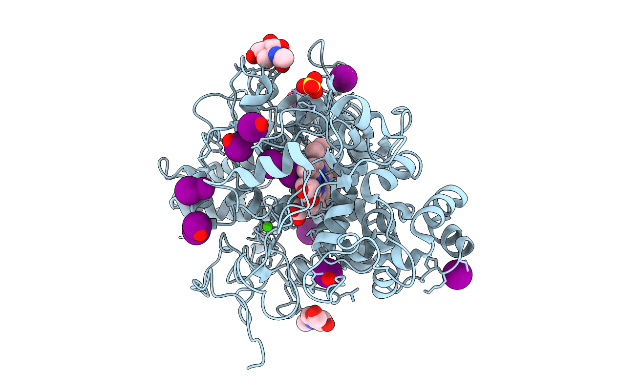
PDB ID:
7VE3
Keywords:
Title:
Structure of the complex of sheep lactoperoxidase with hypoiodite at 2.70 A resolution
Biological Source:
Source Organism(s):
Ovis aries (Taxon ID: 9940)
Expression System(s):
Method Details:
Experimental Method:
Resolution:
2.70 Å
R-Value Free:
0.27
R-Value Work:
0.19
Space Group:
P 1 21 1
