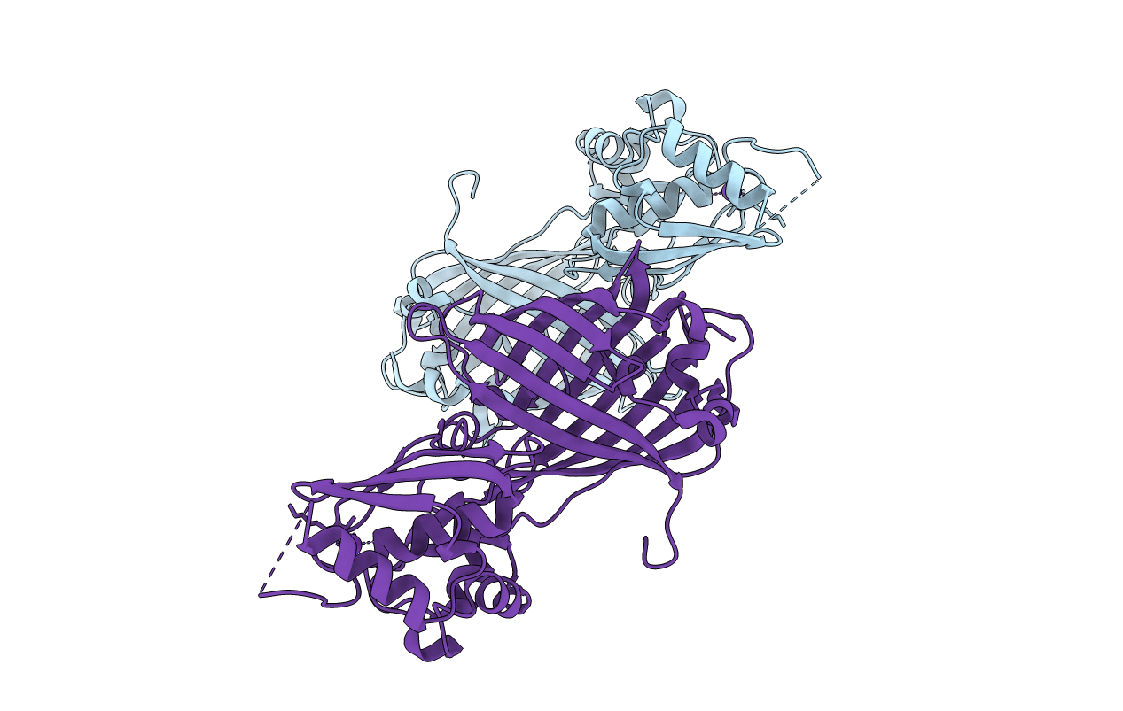
7VCM image
Entry Detail
PDB ID:
7VCM
Title:
crystal structure of GINKO1
Biological Source:
Host Organism:
PDB Version:
Deposition Date:
2021-09-03
Release Date:
2022-07-27
Method Details:
Experimental Method:
Resolution:
1.85 Å
R-Value Free:
0.22
R-Value Work:
0.19
Space Group:
P 1
Macromolecular Entities
Polymer Type:polypeptide(L)
Description:Green fluorescent protein,Potassium binding protein Kbp,Green fluorescent protein
Mutations:F65L,V94I,M304K,V313A,S325G,D330Y,T353V,A356K,H381L
Chain IDs:A, B
Chain Length:388
Number of Molecules:2
Biological Source:Aequorea victoria, Escherichia coli K-12
Modified Residue
Compound ID Chain ID Parent Comp ID Details 2D Image
CRO A SER chromophore
Primary Citation
A sensitive and specific genetically-encoded potassium ion biosensor for in vivo applications across the tree of life.
Plos Biol. 20 e3001772 e3001772 (2022)
PMID: 36067248 DOI: 10.1371/journal.pbio.3001772

Abstact

Potassium ion (K+) plays a critical role as an essential electrolyte in all biological systems. Genetically-encoded fluorescent K+ biosensors are promising tools to further improve our understanding of K+-dependent processes under normal and pathological conditions. Here, we report the crystal structure of a previously reported genetically-encoded fluorescent K+ biosensor, GINKO1, in the K+-bound state. Using structure-guided optimization and directed evolution, we have engineered an improved K+ biosensor, designated GINKO2, with higher sensitivity and specificity. We have demonstrated the utility of GINKO2 for in vivo detection and imaging of K+ dynamics in multiple model organisms, including bacteria, plants, and mice.

Legend

Protein

Chemical

Disease

Primary Citation of related structures