7VB3 image
Entry Detail
PDB ID:
7VB3
Keywords:
Title:
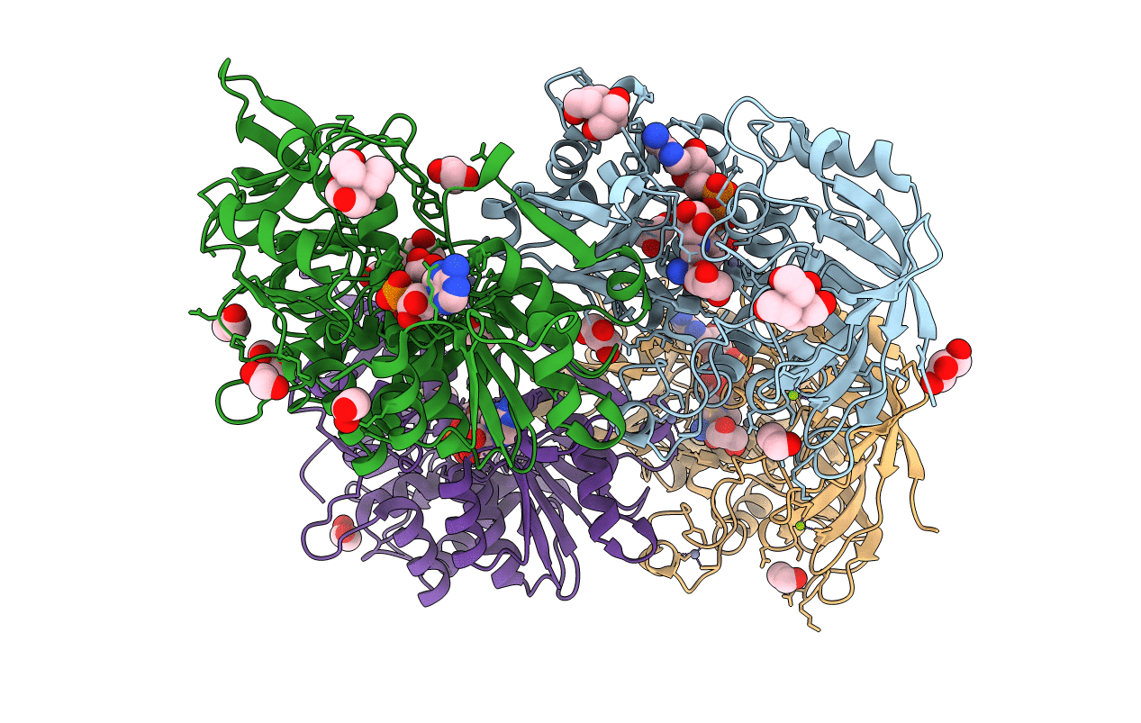
Crystal structure of hydroxynitrile lyase from Linum usitatissimum
Biological Source:
Source Organism:
Host Organism:
PDB Version:
Deposition Date:
2021-08-30
Release Date:
2022-02-09
Method Details:
Experimental Method:
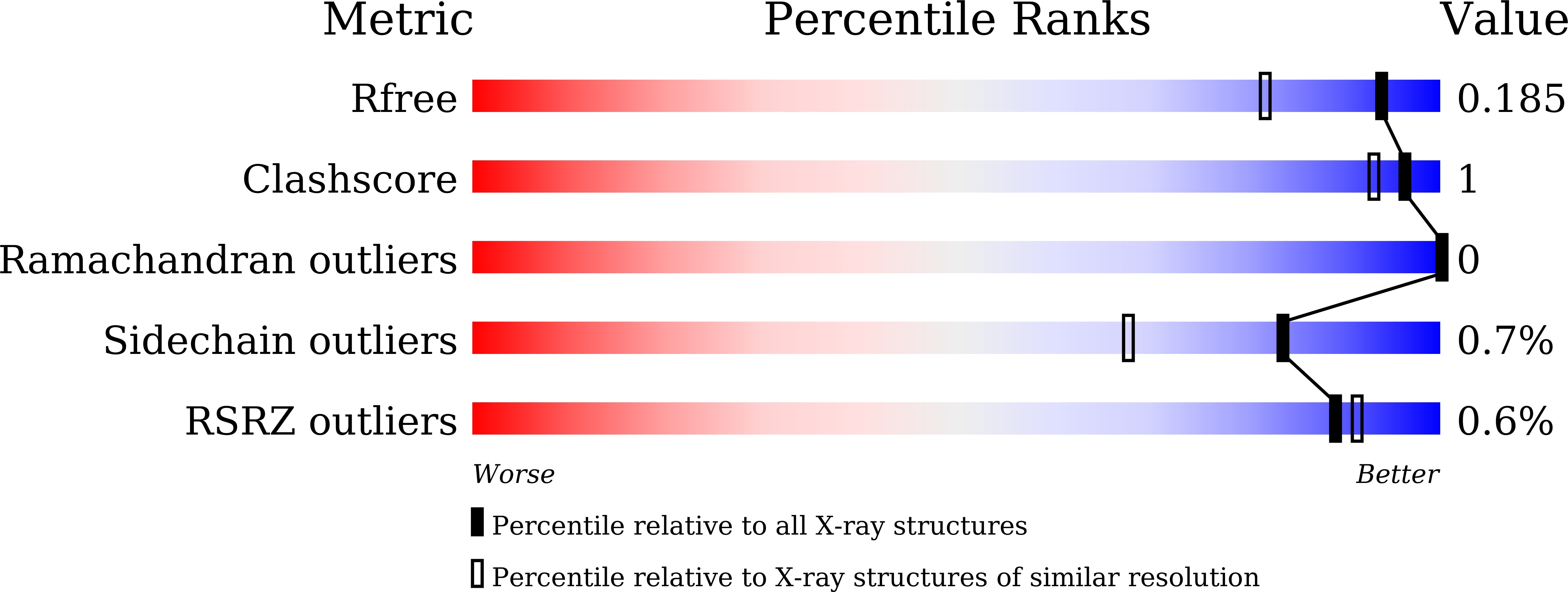
Resolution:
1.48 Å
R-Value Free:
0.18
R-Value Work:
0.15
R-Value Observed:
0.15
Space Group:
P 1 21 1
Macromolecular Entities
Polymer Type:polypeptide(L)
Description:Aliphatic (R)-hydroxynitrile lyase
Chain IDs:A, B, C, D
Chain Length:443
Number of Molecules:4
Biological Source:Linum usitatissimum
Modified Residue
Compound ID Chain ID Parent Comp ID Details 2D Image
SNC A CYS modified residue
Primary Citation
Structural characterization of Linum usitatissimum hydroxynitrile lyase: A new cyanohydrin decomposition mechanism involving a cyano-zinc complex.
J.Biol.Chem. 298 101650 101650 (2022)
PMID: 35101448 DOI: 10.1016/j.jbc.2022.101650

Abstact

Hydroxynitrile lyase from Linum usitatissimum (LuHNL) is an enzyme involved in the catabolism of cyanogenic glycosides to release hydrogen cyanide upon tissue damage. This enzyme strictly conserves the substrate- and NAD(H)-binding domains of Zn2+-containing alcohol dehydrogenase (ADH); however, there is no evidence suggesting that LuHNL possesses ADH activity. Herein, we determined the ligand-free 3D structure of LuHNL and its complex with acetone cyanohydrin and (R)-2-butanone cyanohydrin using X-ray crystallography. These structures reveal that an A-form NAD+ is tightly but not covalently bound to each subunit of LuHNL. The restricted movement of the NAD+ molecule is due to the "sandwich structure" on the adenine moiety of NAD+. Moreover, the structures and mutagenesis analysis reveal a novel reaction mechanism for cyanohydrin decomposition involving the cyano-zinc complex and hydrogen-bonded interaction of the hydroxyl group of cyanohydrin with Glu323/Thr65 and H2O/Lys162 of LuHNL. The deprotonated Lys162 and protonated Glu323 residues are presumably stabilized by a partially desolvated microenvironment. In summary, the substrate binding geometry of LuHNL provides insights into the differences in activities of LuHNL and ADH, and identifying this novel reaction mechanism is an important contribution to the study of hydroxynitrile lyases.

Legend

Protein

Chemical

Disease

Primary Citation of related structures