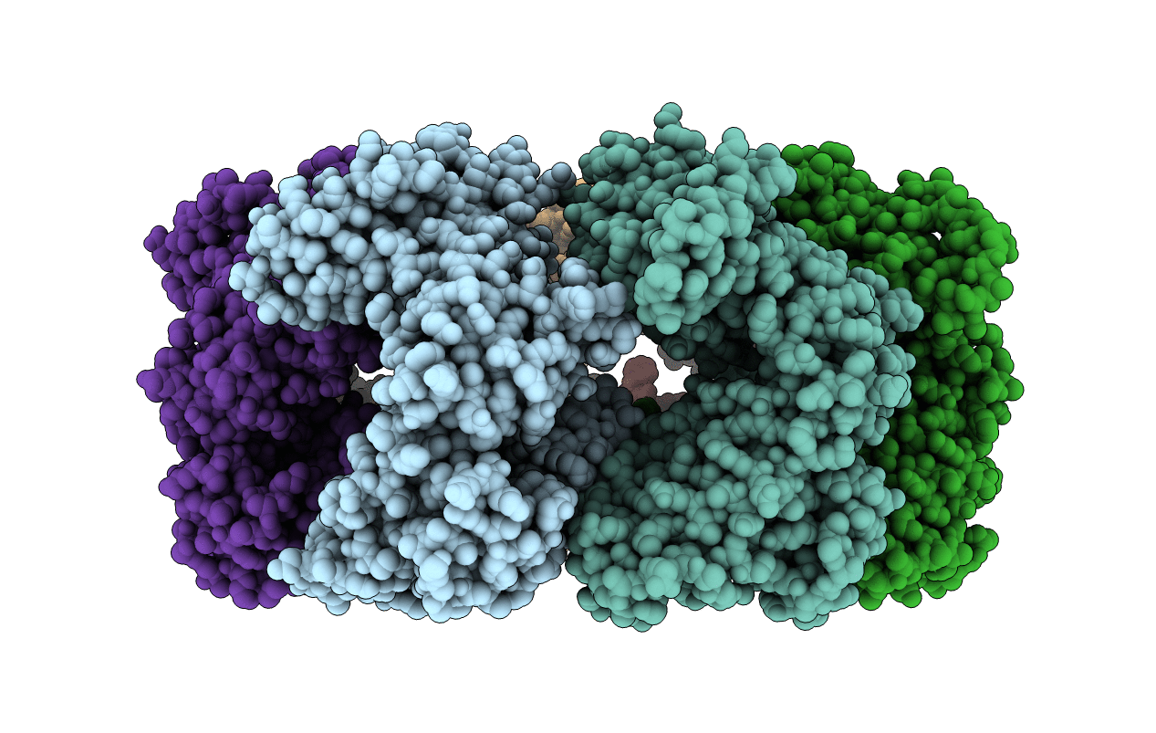
7V9R image
Entry Detail
PDB ID:
7V9R
Keywords:
Title:
Crystal Structure of the heptameric EcHsp60
Biological Source:
Source Organism:
PDB Version:
Deposition Date:
2021-08-26
Release Date:
2022-08-31
Method Details:
Experimental Method:
Resolution:
3.50 Å
R-Value Free:
0.22
R-Value Work:
0.18
R-Value Observed:
0.18
Space Group:
P 21 21 21
Macromolecular Entities
Polymer Type:polypeptide(L)
Description:60 kDa chaperonin
Mutations:M330V
Chain IDs:A, B, C, D, E, F, G
Chain Length:573
Number of Molecules:7
Biological Source:Epinephelus coioides
Ligand Molecules
Primary Citation
Crystal structures of dimeric and heptameric mtHsp60 reveal the mechanism of chaperonin inactivation.
Life Sci Alliance 6 ? ? (2023)
PMID: 36973006 DOI: 10.26508/lsa.202201753

Abstact

Mitochondrial Hsp60 (mtHsp60) plays a crucial role in maintaining the proper folding of proteins in the mitochondria. mtHsp60 self-assembles into a ring-shaped heptamer, which can further form a double-ring tetradecamer in the presence of ATP and mtHsp10. However, mtHsp60 tends to dissociate in vitro, unlike its prokaryotic homologue, GroEL. The molecular structure of dissociated mtHsp60 and the mechanism behind its dissociation remain unclear. In this study, we demonstrated that Epinephelus coioides mtHsp60 (EcHsp60) can form a dimeric structure with inactive ATPase activity. The crystal structure of this dimer reveals symmetrical subunit interactions and a rearranged equatorial domain. The α4 helix of each subunit extends and interacts with its adjacent subunit, leading to the disruption of the ATP-binding pocket. Furthermore, an RLK motif in the apical domain contributes to stabilizing the dimeric complex. These structural and biochemical findings provide new insights into the conformational transitions and functional regulation of this ancient chaperonin.

Legend

Protein

Chemical

Disease

Primary Citation of related structures