Deposition Date
2021-08-23
Release Date
2022-01-05
Last Version Date
2023-11-29
Entry Detail
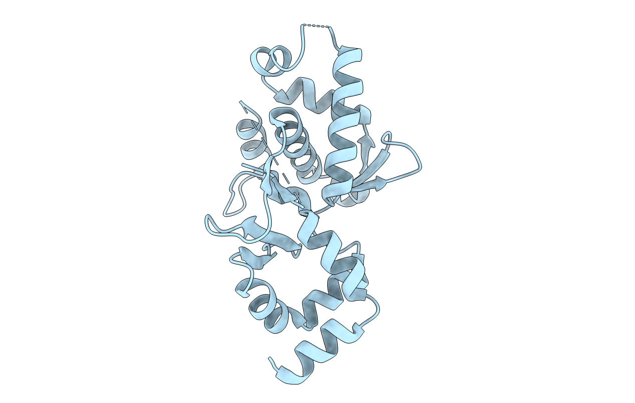
Biological Source:
Source Organism(s):
Shigella flexneri (Taxon ID: 623)
Expression System(s):
Method Details:
Experimental Method:
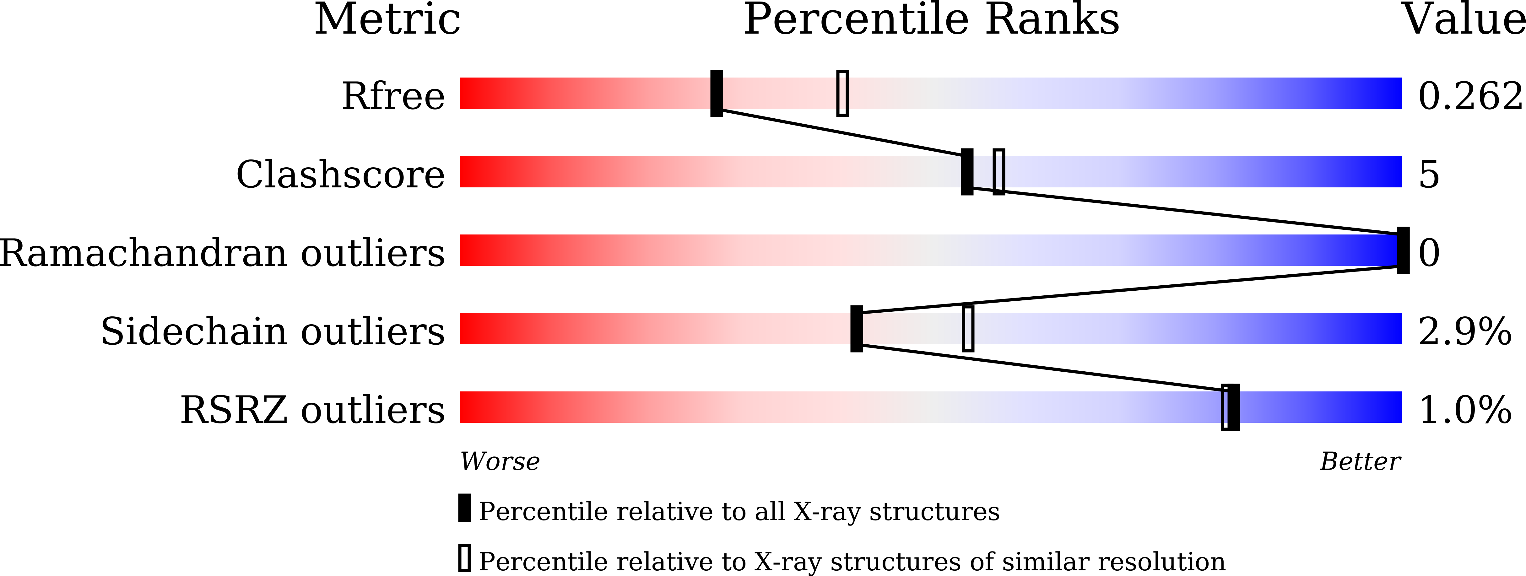
Resolution:
2.44 Å
R-Value Free:
0.26
R-Value Work:
0.21
R-Value Observed:
0.22
Space Group:
C 1 2 1