7V5Y image
Entry Detail
PDB ID:
7V5Y
Keywords:
Title:
Crystal structure of hexameric complex of Sa2YoeB-Sa2YefM toxin-antitoxin from Staphylococcus aureus
Biological Source:
PDB Version:
Deposition Date:
2021-08-18
Release Date:
2021-12-15
Method Details:
Experimental Method:
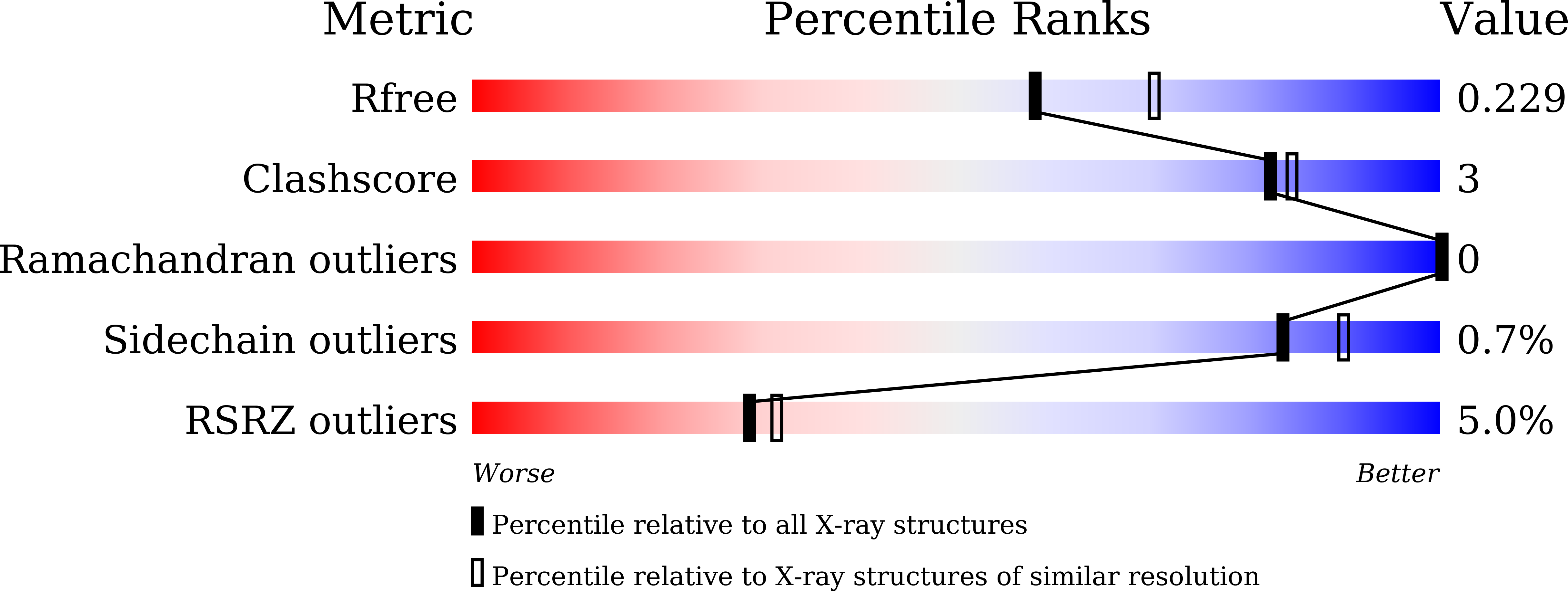
Resolution:
2.25 Å
R-Value Free:
0.22
R-Value Work:
0.18
R-Value Observed:
0.18
Space Group:
P 1 21 1
Macromolecular Entities
Polymer Type:polypeptide(L)
Description:Antitoxin
Chain IDs:A (auth: B), B (auth: D), C (auth: A), D (auth: C)
Chain Length:85
Number of Molecules:4
Biological Source:Staphylococcus aureus (strain NCTC 8325 / PS 47)
Polymer Type:polypeptide(L)
Description:Putative mRNA interferase YoeB
Chain IDs:E, F
Chain Length:88
Number of Molecules:2
Biological Source:Staphylococcus aureus (strain NCTC 8325 / PS 47)
Primary Citation
The two paralogous copies of the YoeB-YefM toxin-antitoxin module in Staphylococcus aureus differ in DNA binding and recognition patterns.
J.Biol.Chem. 298 101457 101457 (2022)
PMID: 34861238 DOI: 10.1016/j.jbc.2021.101457

Abstact

Toxin-antitoxin (TA) systems are ubiquitous regulatory modules for bacterial growth and cell survival following stress. YefM-YoeB, the most prevalent type II TA system, is present in a variety of bacterial species. In Staphylococcus aureus, the YefM-YoeB system exists as two independent paralogous copies. Our previous research resolved crystal structures of the two oligomeric states (heterotetramer and heterohexamer-DNA ternary complex) of the first paralog as well as the molecular mechanism of transcriptional autoregulation of this module. However, structural details reflecting molecular diversity in both paralogs have been relatively unexplored. To understand the molecular mechanism of how Sa2YoeB and Sa2YefM regulate their own transcription and how each paralog functions independently, we solved a series of crystal structures of the Sa2YoeB-Sa2YefM. Our structural and biochemical data demonstrated that both paralogous copies adopt similar mechanisms of transcriptional autoregulation. In addition, structural analysis suggested that molecular diversity between the two paralogs might be reflected in the interaction profile of YefM and YoeB and the recognition pattern of promoter DNA by YefM. Interaction analysis revealed unique conformational and activating force effected by the interface between Sa2YoeB and Sa2YefM. In addition, the recognition pattern analysis demonstrated that residues Thr7 and Tyr14 of Sa2YefM specifically recognizes the flanking sequences (G and C) of the promoter DNA. Together, these results provide the structural insights into the molecular diversity and independent function of the paralogous copies of the YoeB-YefM TA system.

Legend

Protein

Chemical

Disease

Primary Citation of related structures