7UY2 image
Entry Detail
PDB ID:
7UY2
Keywords:
Title:
Structure of RNF31 in complex with FP06649, a Helicon Polypeptide
Biological Source:
Host Organism:
PDB Version:
Deposition Date:
2022-05-06
Release Date:
2022-12-28
Method Details:
Experimental Method:
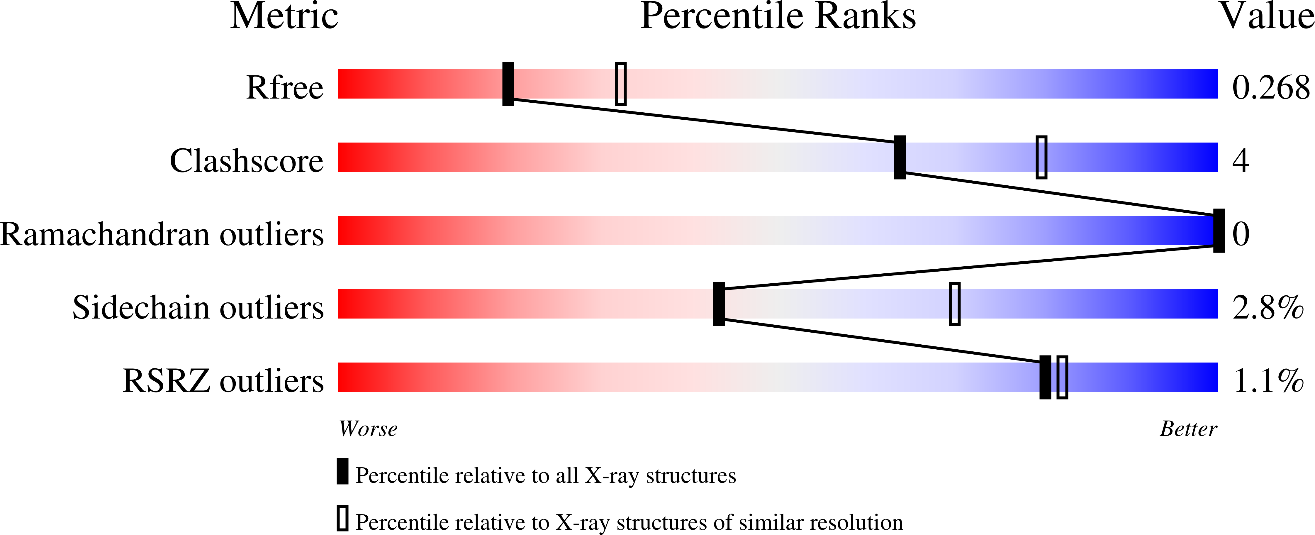
Resolution:
2.51 Å
R-Value Free:
0.27
R-Value Work:
0.21
R-Value Observed:
0.21
Space Group:
P 61
Macromolecular Entities
Polymer Type:polypeptide(L)
Description:E3 ubiquitin-protein ligase RNF31
Chain IDs:A, B
Chain Length:184
Number of Molecules:2
Biological Source:Homo sapiens
Polymer Type:polypeptide(L)
Description:Helicon FP06649
Chain IDs:C, D
Chain Length:14
Number of Molecules:2
Biological Source:synthetic construct
Primary Citation
De novo mapping of alpha-helix recognition sites on protein surfaces using unbiased libraries.
Proc.Natl.Acad.Sci.USA 119 e2210435119 e2210435119 (2022)
PMID: 36534810 DOI: 10.1073/pnas.2210435119

Abstact

The α-helix is one of the most common protein surface recognition motifs found in nature, and its unique amide-cloaking properties also enable α-helical polypeptide motifs to exist in membranes. Together, these properties have inspired the development of α-helically constrained (Helicon) therapeutics that can enter cells and bind targets that have been considered "undruggable", such as protein-protein interactions. To date, no general method for discovering α-helical binders to proteins has been reported, limiting Helicon drug discovery to only those proteins with previously characterized α-helix recognition sites, and restricting the starting chemical matter to those known α-helical binders. Here, we report a general and rapid screening method to empirically map the α-helix binding sites on a broad range of target proteins in parallel using large, unbiased Helicon phage display libraries and next-generation sequencing. We apply this method to screen six structurally diverse protein domains, only one of which had been previously reported to bind isolated α-helical peptides, discovering 20 families that collectively comprise several hundred individual Helicons. Analysis of 14 X-ray cocrystal structures reveals at least nine distinct α-helix recognition sites across these six proteins, and biochemical and biophysical studies show that these Helicons can block protein-protein interactions, inhibit enzymatic activity, induce conformational rearrangements, and cause protein dimerization. We anticipate that this method will prove broadly useful for the study of protein recognition and for the development of both biochemical tools and therapeutics for traditionally challenging protein targets.

Legend

Protein

Chemical

Disease

Primary Citation of related structures